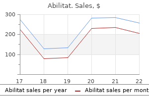
Proven 15 mg abilitat
Detection is enhanced by culture on rabbit or human blood agar rather than on extra generally used sheep blood agar due to larger colony measurement and wider zones of hemolysis utter depression definition abilitat 15 mg generic line. Two biotypes of A haemolyticum have been recognized: a tough biotype predominates in respiratory tract infections and a easy biotype is mostly associated with skin and softtissue infections depression kanji 10 mg abilitat purchase with mastercard. Treatment Erythromycin is the drug of selection for treating tonsillopharyngitis attributable to A haemolyticum. A haemolyticum is also prone in vitro to azithromycin, clindamycin, cefuroxime, vancomycin, and tetracycline. Failures in therapy of pharyngitis with penicillin have been reported, perhaps due to this intracellular residing pathogen. In disseminated an infection, parenteral penicillin plus an aminoglycoside could additionally be used initially as empirical therapy. A haemolyticum appears strongly gram-positive in young cultures however turns into more gramvariable after 24 hours of incubation as in this photograph. During the larval migratory part, an acute transient pneumonitis (L�ffler syndrome) associated with fever and marked eosinophilia can occur. Children are prone to this complication due to the small diameter of the intestinal lumen and their propensity to acquire massive worm burdens. Worm migration could cause peritonitis secondary to intestinal wall perforation and common bile duct obstruction leading to biliary colic, cholangitis, or pancreatitis. Adult worms may be stimulated to migrate by tense conditions (eg, fever, illness, or anesthesia) and by some anthelmintic medicine. Female worms produce roughly 200,000 eggs per day, that are excreted in stool and should incubate in soil for 2 to three weeks for an embryo to turn into infectious. Following ingestion of embryonated eggs, normally from contaminated soil, larvae hatch within the small intestine, penetrate the mucosa, and are transported passively by portal blood to the liver and lungs. Infection with A lumbricoides is most typical in resourcelimited nations, together with rural and urban communities characterized by poor sanitation. Incubation Period Approximately 8 weeks (interval between ingestion of eggs and improvement of egglaying adults). Diagnostic Tests Ova routinely are detected by examination of a recent stool specimen using gentle microscopy. Infected people also could pass adult worms from the rectum, from the nose after migration through the nares, and from the mouth, usually in vomitus. Treatment Albendazole (taken with meals in a single dose), mebendazole for three days, or ivermectin (taken on an empty abdomen in a single dose) are recommended for remedy of ascariasis. In 1-year-old youngsters, the World Health Organization recommends reducing the albendazole dose to half of that given to older children and adults. Reexamination of stool specimens 2 weeks after therapy to decide whether the worms have been eliminated is helpful for assessing effectiveness of therapy. Surgical intervention occasionally is critical to relieve intestinal or biliary tract obstruction or for volvulus or peritonitis secondary to perforation. This worm is a feminine, as evidenced by the size and genital girdle (the darkish round groove at bottom area of image). After infective eggs are swallowed (4), the larvae hatch (5), invade the intestinal mucosa, and are carried by way of the portal, then systemic circulation to the lungs (6). The larvae mature additional in the lungs (10�14 days), penetrate the alveolar partitions, ascend the bronchial tree to the throat, and are swallowed (7). Invasive aspergillosis happens nearly exclusively in immunocompromised sufferers with extended neutropenia (eg, cytotoxic chemotherapy), graft-versus-host disease, or impaired phagocyte perform (eg, chronic granulomatous disease, immunosuppressive remedy, corticosteroids). Children at highest risk include children with new-onset or a relapse of hematologic malignancy and allogeneic hematopoietic stem cell transplant recipients. The hallmark of invasive aspergillosis is angioinvasion with ensuing thrombosis, dissemination to other organs and, sometimes, erosion of the blood vessel wall with catastrophic hemorrhage. Aspergillomas and otomycosis are 2 syndromes of nonallergic colonization by Aspergillus species in immunocompetent youngsters. Allergic bronchopulmonary aspergillosis is a hypersensitivity lung disease that manifests as episodic wheezing, expectoration of brown mucus plugs, low-grade fever, eosinophilia, and transient pulmonary infiltrates. Allergic sinusitis is a far much less frequent allergic response to colonization by Aspergillus species than is allergic bronchopulmonary aspergillosis. Aspergillus fumigatus is the most typical cause of invasive aspergillosis, with Aspergillus flavus being the next most common. Several other species, including Aspergillus terreus, Aspergillus nidulans, and Aspergillus niger, additionally trigger invasive human infections. Epidemiology the principal route of transmission is inhalation of conidia (spores) originating from a number of environmental sources (plants, vegetables, mud from building or demolition), soil, and water supplies (eg, bathe heads). Incidence of disease in transplant recipients is highest during periods of neutropenia or throughout treatment for graft-versus-host disease. Health care�associated outbreaks of invasive pulmonary aspergillosis in vulnerable hosts have occurred in which the probable source of the fungus was a close-by building site or defective ventilation system. Diagnostic Tests Dichotomously branched and septate hyphae, recognized by microscopic examination of 10% potassium hydroxide moist preparations or of Gomori methenamine silver nitrate stain of tissue or bronchoalveolar lavage specimens, are suggestive of the diagnosis. Aspergillus species could be a laboratory contaminant, but when evaluating results from sick, immunocompromised patients, restoration of this organism frequently indicates an infection. An enzyme immunosorbent assay serologic take a look at for detection of galactomannan, a molecule discovered within the cell wall of Aspergillus species, is available commercially and has been found to be useful in kids and adults. Monitoring of serum antigen concentrations twice weekly in periods of highest danger (eg, neutropenia and energetic graft-versus-host disease) may be helpful for early detection of invasive aspergillosis in at-risk sufferers. Falsepositive test outcomes have been reported and could be associated to consumption of meals merchandise containing galactomannan (eg, rice and pasta) or from cross-reactivity with antimicrobial brokers derived from fungi (eg, penicillins). In allergic aspergillosis, analysis is suggested by a typical scientific syndrome with elevated whole concentrations of immunoglobulin (Ig) E (1,000 ng/mL) and Aspergillusspecific serum IgE, eosinophilia, and a positive end result from a skin check for Aspergillus antigens. In children with cystic fibrosis, the prognosis is tougher, as a outcome of wheezing and eosinophilia not related to allergic bronchopulmonary aspergillosis usually are current. Treatment Voriconazole is the drug of selection for invasive aspergillosis, except in neonates, for whom amphotericin B deoxycholate in excessive doses is beneficial. Monitoring of serum galactomannan serum concentrations twice weekly may be helpful to assess response to therapy concomitant with medical and radiologic analysis. Caspofungin has been studied in pediatric sufferers older than three months as salvage remedy for invasive aspergillosis. Itraconazole alone is an alternative for gentle to moderate cases of aspergillosis, although extensive drug interactions and poor absorption (capsular form) restrict the utility of itraconazole. Lipid formulations of amphotericin B could be thought-about, however A terreus is immune to all amphotericin B merchandise. Decreasing immunosuppression, if potential, particularly decreasing corticosteroid dose, is necessary to illness control.
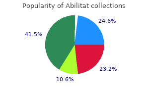
Purchase 20 mg abilitat with amex
It has been instructed that elective oophorectomy on the time of hysterectomy might forestall 10% of ovarian cancers depression research purchase 20 mg abilitat otc,fifty three anxiety meds discount abilitat 15 mg mastercard,54 though curiously, hysterectomy alone appears to cut back the danger of subsequent ovarian malignancy. Additionally, ovarian operate could be affected by surgical interruption to the uterine blood supply. Indeed, hysterectomy with ovarian conservation has been associated with premature ovarian failure. As properly as the risk of subsequent ovarian malignancy, conserving ovarian tissue could be associated with ongoing morbidity. Residual ovary syndrome is a explanation for pelvic pain and dyspareunia, and ends in additional surgery in 1�2% of cases following hysterectomy. The cumulative ovar- 147 Section B Benign Conditions: the Cervix, Vagina and Vulva, Uterus, Ovaries and Fallopian Tubes Table 9. Psychological Many women quite rightly regard the ovary as the gland that chiefly determines their femininity. Regardless of age and menopausal standing, some women may give consent to hysterectomy solely with the proviso that their ovaries may be retained. In view of the above, sure guiding rules should be followed: Before the menopause as a lot as the age of fifty, wholesome ovaries should normally be conserved. Risk-reducing salpingooophorectomy is the best preventative measure for ovarian or tubal cancer in high-risk women, though a four. In circumstances of prolapse, oophorectomy may be achieved fairly simply by the vaginal route. In other instances of vaginal hysterectomy, where entry is restricted, laparoscopic assistance may be used or endoscopic instrumentation through the open vaginal vault. The standard historic practice of elective oophorectomy at abdominal hysterectomy in ladies over the age of 45 is now not acceptable, not only in view of the confirmed advantages of functioning ovarian tissue within the majority of women, but also the supply of laparoscopic surgical procedure means most subsequent benign ovarian circumstances may be handled without major belly surgery. Endocrine Following bilateral oophorectomy, oestrogen lack could trigger extreme and severe well being issues: Cardiovascular: Low oestrogen levels end in modifications in lipid and carbohydrate metabolism, and insulin sensitivity, which can predispose to cardiovascular disease. Osteoporosis: After the menopause, there is a rise in bone loss with as much as 5% of trabecular and 1. Vasomotor: the signs of the climacteric similar to scorching flushes and night sweats are said to be extra severe after a surgical menopause. Libido: Reduction in circulating androgens could additionally be responsible for a lack of libido. The evidence for pre-treatment in the case of dysfunctional uterine bleeding or endometriosis is less clear. In the case of an enormous fibroid uterus extending above the umbilicus, it could also allow for adequate shrinkage to obtain hysterectomy via a Pfannenstiel somewhat than a midline belly incision. Prophylactic Antibiotics the advent of antibiotic use in the administration of postoperative sepsis after hysterectomy significantly lowered morbidity and mortality charges. Antibiotic prophylaxis is believed to scale back postoperative infections by lowering the variety of contaminant organisms and rendering tissue fluid less appropriate as a tradition medium. A number of regimens have been proven to be beneficial in lowering pelvic infection and febrile morbidity at vaginal hysterectomy. For the needs of this textual content we describe conventional dissection and ligation of pedicles using absorbable suture materials. There are now a wide range of electrosurgical instruments and staple gadgets which were developed for open surgical procedure, which are able to sealing large vessels successfully. The Trendelenburg place is useful, although not all the time essential, and the anaesthetist should guarantee sufficient muscle rest to forestall increased intra-abdominal stress that may pressure the intestines into the operative area. The Ovarian and Round Ligament Pedicles: the stomach is opened in the traditional way with the affected person mendacity within the Trendelenburg position. Self-retaining retractors are launched and the intestines packed off in the event that they protrude into the operation area. The uterus is drawn out of the stomach wound and over to the left facet of the patient so that the best tube and ovary become visible. Particular attention must be paid to obtaining full haemostasis in the area of ovaries. Spencer Wells) is positioned over the best ovarian ligament and the right Fallopian tube, and a second is positioned on the right round ligament. Spencer Wells forceps are additionally applied on the uterine aspect of each of those clamps and the intervening tissues reduce through with scissors. If the clamps have been correctly placed, there shall be no bleeding from the minimize tissues, however sometimes a tortuous terminal department of the uterine artery might escape the medial clamp and need to be caught up by an extra pair of artery forceps. The clamp placed over the ovarian ligament and the Fallopian tube must be positioned as close to to the uterus as possible as a end result of when the tissues enclosed by the clamp are tied, the ligature materials may cut by way of the tissue of the ovarian ligament and result in troublesome bleeding. Furthermore, secondary haemorrhage after the operation is regularly due to vessels that retract away from the ligatures positioned around the tissues enclosed by this clamp. In such instances, little can be carried out to mobilise the uterus till the endometriotic disease has been excised or parametrial tissues have been divided. Dividing the Anterior Leaf of the Broad Ligament: the uterus is firmly pulled to the left side of the affected person by the assistant, and the above process repeated. Then, with dissecting forceps and scissors, starting from the cut round ligament, the surgeon divides the peritoneum, which forms the anterior leaf of the broad ligament, downwards and inwards in path of the uterovesical pouch. If the round ligament is now drawn laterally by an assistant, a thin sheet of endopelvic fascia will be seen passing downwards from the round ligament in the path of the lateral border of the uterus. This sheet of fascia, although thin, is all the time present, and it reaches the uterus anterior to the uterine vessels as they cross along its facet. It is the follow of the authors at all times to minimize via this fascial layer downwards and forwards 9. Alternatively, the uterus may be manipulated by Spencer Wells forceps placed at every cornu. In entrance, the upper restrict of the bladder could be identified, while on the anterior surface of the uterus is a V-shaped fossa. In this fashion the uterine vessels are cleanly uncovered, or skeletonised, and in a sub-total hysterectomy they are often clamped with a curved clamp low down close to the junction of the body with the cervix. In a total hysterectomy, the clamping and division of the uterine pedicle should be postponed till the bladder has been freed from the cervix and dislocated downwards, as described in the next steps of the operation. Reflecting the Bladder Inferiorly: To obtain bladder reflection, the peritoneum of the uterovesical pouch must first be divided. This surgical airplane could be less obvious if there has been previous dissection of the uterovesical area. In the normal uterus, the peritoneum is reflected onto the anterior surface of the uterus from the bladder roughly on the stage of the internal os. If the anterior peritoneal floor of the uterus is examined, a small fossa can all the time be identified just above the level of the inner os, and quite regularly a small ridge could be identified on all sides of the uterus, which passes from the decrease fringe of this fossa within the midline upwards and laterally. The peritoneum is adherent to the uterus over this fossa, however instantly under it can easily be separated from the underlying constructions.
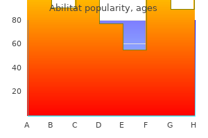
Discount abilitat 20 mg on line
A lateral strand also separates the perianal area from the ischiorectal fossa9 with a forward extension tethering the interior anal sphincter by way of attachment of the longitudinal muscle to the perineal membrane on the apex of the perineal physique mood disorder treatment center buy 15 mg abilitat amex. This tethering extends parasagittally and may be liable for childbirth tears of the internal anal sphincter without essentially the exterior sphincter being fully divided depression test german abilitat 15 mg order on line. Pelvic Fascia and Endopelvic Fascia the pelvic fascia is defined because the fascial tissue which covers the upper and lower surfaces of the levator ani muscles, together with the medial surfaces of the two obturator internus muscular tissues. Between the pelvic fascia and the peritoneum above, all the unfastened tissues are greatest referred to because the endopelvic fascia. In addition, the vagina and cervix have their very own fascial layer and the same remarks apply to the rectum, the bladder and the urethra. Furthermore, each of these is attached to adjacent organs and the pelvic facet partitions by pelvic fascia which may be recognised at operation. In the anterior compartment, this fascia is hooked up to a tendinous arc simply medial to the attachment of the levator ani muscle to its personal tendinous arc (white line) which is on the covering of the obturator internus muscle. Tears of the endo-pelvic fascia are the initiating event for numerous kinds of vaginal prolapse. There can additionally be thin muscularis mucosae within the subepithelial house above the dentate line. A condensation of the bottom portion of the inside circular layer types the interior anal sphincter. The rugose projections on the anterior vaginal wall lie beneath this level, and intervening between the vaginal 18. Care wall and the fascia covering the urethra is cavernous tissue, which have to be cut through with a scalpel when the vaginal wall is dissected from the fused vaginal and urethral fascia. The fused vaginal and urethral fascia forms a condensation of tissue which is connected laterally to every pubic ramus and extends from the bladder wall or urethrovesical junction to the urethral meatus. The ligamentous supports of the urethra, the place it passes beneath the pubic arch, have been termed the anterior and posterior pubo-urethral ligaments. Between these supports, and arising by the origin of the levator ani muscle tissue on the physique of the pubis, may be found the extrinsic rhabdosphincter or compressor urethrae. When the pre-rectal fascia has been torn in circumstances of rectocoele, the anterior wall of the rectum presents as a layer of muscle barely corrugated longitudinally with outstanding vessels. This fascial layer has to be penetrated to gain access to the sacrospinous ligament for sacrospinous colpopexy operations. Immediately beneath the peritoneum, passing from the bladder to the uterus is a skinny layer of tissue, the vesicocervical ligament. If during a vaginal operation the cervix is pulled down and the restrict of the bladder uncovered the same bands of tissue may be seen to cross from the bladder to the cervix. Three major condensations could be recognised; one is located within the midline, while two lie laterally. The lateral condensations beneath the bladder pillars are the pubocervical ligaments which are the anterior limb of the transverse cervical or cardinal ligaments. The pubocervical ligaments are answerable for retaining the cervix and higher vagina inside the anterior compartment of the pelvis. If attenuated or destroyed, they allow backward rotation of the entire vagina into the hole of the sacrum (retrocession), a displacement which has to be distinguished from anterior vaginal wall prolapse (cystocoele), with which nevertheless it could be associated. The posterior arcs of the cardinal ligaments are the uterosacral ligaments which embrace the rectum and the pouch of Douglas. Moreover, when traced downwards and inwards it leads to the bladder pillar which may then be identified. The roof of the canal is formed by connective tissue which surrounds the uterine artery, whereas under the ureteric canal lies the principle portion of the cardinal ligament. The paravesical house lies in front of every cardinal ligament and the pararectal space behind. These areas could be recognised at operation and are easily opened up as they contain only very tenuous mobile tissue condensations. To dissect in a aircraft posterior to this 20 Surgical Anatomy fascia is to invite major haemorrhage. There is a useful airplane of loose areolar tissue immediately behind the rectum however in front of this fascia. The urogenital hiatus is inevitably stretched or torn in childbirth and will never return to its pristine state. Much of the pelvic help offered to the pelvic contents on this massive delivery canal is dynamic. Pelvic muscular leisure thus results in undue stress and stretch on the fastened ligamentous and fascial helps of the uterus and vagina, which, in flip offers rise to prolapse. Discrepancies may also be discovered within the descriptions in anatomical textbooks of the structure of the perineal physique and of the sphincter muscles of the anus. Dissecting room subjects are of limited worth for anatomical researches, as fixation, and even anaesthesia, can alter the connection between muscular components, such because the exterior and inside anal sphincters. Modem imaging (endosonography and magnetic resonance) has helped to elucidate the area, however images have had to be validated by serial dissection and histological confirmation earlier than interpretation can be secure. Posterolaterally, the origin is from the tendinous arc (white line) over the obturator internus muscle as far as the ischial backbone. The pubococcygeus muscles are inserted into the coccyx and into the anococcygeal raphe. The puborectalis muscle, which is functionally crucial, actually types a sling across the anorectal junction from the body of 1 pubic bone to the opposite. Because of its firm attachment to the lateral vaginal wall, it acts functionally as a vaginal sphincter without truly encircling that organ. In spite of this anatomical reality, if the affected person is suffering from posterior vaginal wall prolapse and her levator muscular tissues are divaricated, and the tissues which normally bind together the two levator muscular tissues between the vagina and the rectum are either stretched or torn through, not only can a rectocoele be controlled if the two levator muscles are artificially introduced together between the vagina and the rectum, but additionally anal sphincter operate may be improved. The nerve supply to the levator ani muscle reaches the muscle on its visceral (pelvic) aspect. In the female, the perineal membrane which covers the space beneath the inferior pubic rami is successfully divided into three parts by the vagina. The related superficial and deep perineal pouches could also be discovered lateral to the lowest part of 21 Section A Introduction, Anatomy, Pre-op. Care the vagina, where the two halves of the perineal membrane itself present lateral assist. The superficial transverse perineal muscle overlies the free posterior border of the perineal membrane and decussates with the superficial external anal sphincter. The deep transverse perineal muscles occupy a pouch adjacent to the perineal membrane on either side of the vagina. Their medial margin is near the attachment of the puborectalis to the lateral wall of the vagina. Anteriorly, inside the subcutaneous tissue of the vestibule may be found fibres of the bulbospongiosus muscle which is commonly deficient within the midline and due to this fact not identified in a median episiotomy. It is a weak introital sphincter however the fibres could typically be discovered in the pedicle of fats developed for the Martius operation (see Chapter 21).
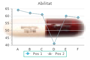
15 mg abilitat discount free shipping
In addition depression kurze definition 20 mg abilitat order fast delivery, the bladder could additionally be drawn up high onto the anterior surface of the uterus and be traumatised when the peritoneum in front of the uterus is being incised depression symptoms on dogs generic abilitat 10 mg online. As with all hysterectomies, every effort should be made to discover the proper tissue plane within the uterovesical peritoneal fold. Broad ligament fibroids: the uterine vessels lie alongside the aspect of the physique of the uterus. Fibroids increasing laterally into the broad ligament rarely displace these vessels. Therefore, if a fibroid grows out from the uterus into the broad ligament, it lies either anterior or posterior to the vessels. Fibroids posterior to the uterine vessels can extend to a point along the uterosacral ligaments. Broad ligament fibroids are often giant sufficient to almost fully fill the pelvis. If they arise from the decrease part of the uterus, they have an inclination to displace the ureter upwards and outwards. The ureter is sometimes firmly related to the capsule of the fibroid, and it must be dissected clear. The bladder is commonly displaced upwards and forwards with the end result that the angle of the bladder along with the ureter lies at a much higher degree than normally. It may be essential in such cases to divide the uterine vessels comparatively high up and to separate them on their medial aspect from the uterus earlier than dissecting downwards into the pelvis to reach either the cervix or the vagina. Occasionally, the stretched ureter can resemble a hypertrophied uterine artery, and if doubtful, the ureter ought to be traced downwards from the purpose where it crosses the pelvic brim. The bladder, the ureter and the uterine vessels are retracted laterally, and the parametrial tissues clamped and divided. However difficult the case, the surgeon can work with confidence offered that the ureter is recognized on either side along with the higher part of the bladder. Intra-operative myomectomy: In some cases with large uterine fibroids, it can be useful to enucleate the fibroid from its capsule intra-operatively earlier than making an attempt to perform a hysterectomy. This is simple to perform and might significantly scale back the volume of the uterus, allowing the surgeon higher freedom of motion and anatomical exposure in the depths of the pelvis. In the case of a cervical fibroid, myomectomy could additionally be achieved by sagittal hemisection of the smaller uterine body. The obstructing tumour is shelled out in a matter of moments, and the capsule and mattress can then be rendered relatively cold by the application of several vulsella to the bleeding surfaces. An in any other case tough hysterectomy instantly becomes simplified, typically with the need for a smaller belly incision and with a discount in subsequent operating time. Endometriosis: the management of endometriosis is mentioned intimately in Chapter eleven. The surgical aim is to take away all endometriotic and adenomyotic illness, within the hope of reaching resolution of symptoms. Removal of the ovaries, the cervix and another endometriotic disease is subsequently advocated at hysterectomy. The primary surgical concern is loss of tissue planes and altered anatomy because of the disease course of. In more extreme cases of rectovaginal endometriosis, resection of a disc or even a section of enormous bowel, with subsequent end-to-end re-anastomosis, may be warranted in order to clear the pelvis of endometriotic disease. Congenital uterine abnormalities: these are discussed in more element in Chapter eight. Similar concerns apply bilaterally when the uterine physique is completely bifid (uterus didelphys). The pre-operative preparation of the affected person is the same as that employed for different vaginal operations for prolapse. The affected person is placed within the lithotomy place, the vagina and vulva disinfected and the surgical subject surrounded with sterile drapes. If the prolapse is of a extreme diploma, it may not be needed for the assistants to make use of lateral vaginal retractors. Vaginal Incision and Demarcation of Lateral Vaginal Flaps: A midline or inverted V incision is made in the anterior vaginal wall in exactly the identical means as if for an anterior colporrhaphy. At the cervical end, the incision is then continued laterally at right angles to the unique incision and continued across the cervix so as to full the circumcision of the cervix. Distortion of the anatomy inside the base of the uterus and broad ligaments because of a big cervical fibroid could make elimination of the uterus hazardous. Surgical safety and operative time can be improved by preliminary enucleation of the fibroid. The Uterus the vaginal flaps are separated from the bladder within the vesicovaginal house, while near the urethra the flaps are dissected clear of the posturethral ligament with a scalpel. Freeing the Bladder Upwards to Expose and Open the Uterovesical Pouch: the peritoneal cavity may be entered by both an anterior or posterior colpotomy. In the case of prolapse, an anterior colpotomy is normally advocated, although that is depending on the operator. The peritoneum is split on both sides as far laterally as is possible using a lateral stretching motion with both index fingers. Excision of Redundant Vaginal Flaps: the redundant lateral vaginal flaps can now be excised, removing both vaginal wall and vaginal fascia. The cervix is elevated to allow the incision to be completed across the posterior fornix. Opening the Pouch of Douglas: the assistant now pulls the cervix upwards and forwards, and the surgeon shows the pouch of Douglas by exerting traction on the vaginal flap of the posterior fornix in a downward course. The peritoneum of the pouch of Douglas is now opened and the incision prolonged laterally on either side so far as the dense fibres of the uterosacral ligaments. If bowel presents, it may be changed, and a wet gauze pack (attached to identification forceps) inserted. The second of these transfixion sutures is left lengthy and secured without rigidity to the operation towels as a marker. Clamping and Dividing the Main Pedicles: the surgeon now proceeds from below (inferiorly) upwards along the tissues of the broad ligament by clamping, chopping and ligating each chew. A longitudinal incision has been made within the anterior vaginal wall together with a transverse incision. The diagram shows the strategy of opening the vesicovaginal space by dissecting the vagina together with the vaginal fascia away from the bladder. The cervix is pulled upwards and forwards, inserting the uterosacral ligaments on the stretch. The vaginal flaps which were dissected from the cervix are drawn posterolaterally.
15 mg abilitat order
The inter-iliac nodes are then approached through a normal parietal incision between the spherical and infundibulo-pelvic ligaments great depression relief definition generic abilitat 10 mg mastercard. The paravesical area is opened to expose the exterior iliac vein extending inferiorly to the obturator nerve medial to the obturator internus muscle anxiety scale discount abilitat 20 mg on line, which is properly visualised. The nodebearing tissue is dissected free in a cranial course, exposing the obliterated umbilical artery medially and the external iliac vessels laterally, the dissection being carried to the bifurcation of the frequent iliac arteries. This is probably adequate for diagnostic functions, but for therapeutic lymphadenectomy it might be acceptable to extend the dissection cranially to the decrease lumbar (para-aortic) nodes. It ought to be remembered that lymphatics accompanying the ovarian vessels drain to lumbar nodes at the stage of the renal vessels. Diagnostic lymphadenectomy subsequently has a place to be able to decide whether radical surgery or radiotherapy ought to be chosen to treat cervical cancer. Laparoscopic Radical Hysterectomy It is now apparent that laparoscopic strategies may be used for carrying out a radical hysterectomy utilizing the same rules and steps because the classical procedure. Since the technique was first launched some 20 years in the past,19 great advances have been made with improved equipment and more lately the introduction of robotic surgical procedure. The number of lymph nodes obtained will increase with operator experience and 310 Uterus and Cervix Cancer unnecessary surgical steps, has proven that operative instances could additionally be shortened while on the identical time pelvic and para-aortic node dissection methods could also be improved with an increasing and satisfactory nodal rely achieved. An enchancment in surgical efficiency has been demonstrated with rising experience. Whilst operating time could additionally be longer initially in robotic cases, clinical outcomes are related for both robotic assisted and laparoscopic hysterectomy, and blood loss seems less for these undergoing surgical procedure by robotic help. The shorter studying curve implies that the typical surgeon will master the robotic method extra speedily than pure laparoscopic procedures. The main scope for therapeutic lymphadenectomy must be in support of radical vaginal hysterectomy or functionpreserving procedures similar to radical trachelectomy. The Schauta operation went into disfavour because of inaccessibility of the lymph nodes and rising bladder morbidity. Pelvic lymphadenectomy is carried out initially identifying tissue plains and spaces. The exterior iliac vein and artery are separated from the pelvic walls allowing identification of the exterior iliac lymph nodes in addition to the obturator nodes. The pararectal house is developed and the uterine artery both clipped or cauterised close to its origin on the anterior division of the internal iliac artery. The ureteric tunnel is opened and the dissection continued to obtain sufficient parametrial and paracervical tissue precisely as for an open radical hysterectomy. Nerve-sparing surgical procedure is feasible and extra profitable by direct visualisation of the lateral pelvic and splanchnic nerves. If the procedure is to be accomplished by the vaginal route then the cuff of vagina is divided and mobilised vaginally but alternatively the vagina may be incised as for the standard belly process. The specimen could then be removed by the vaginal route and subsequently the vaginal vault closed utilizing steady 2/0 vicryl. The V lock suture is particularly helpful for this ensuring safer haemostasis with out the chance of sutures slipping. Long-Term Complications of Radical Pelvic Surgery and Their Prevention the major long-term morbid sequel of radical hysterectomy is impairment of bladder function. Peri-vesical scarring may significantly cut back bladder capacity (non-compliant bladder) and, finally, broad lateral parametrial dissection and division of the cardinal ligaments may transect the pelvic nerves and thus denervate the parasympathetic motor supply to the bladder. A lesser degree of nerve trauma (neuropraxia) may get well however have the opposite impact of manufacturing inappropriate detrusor muscle activity (detrusor instability). Identification and preservation of the hypogastric and lateral pelvic nerve plexuses will help to scale back this bladder and rectal dysfunction. It is necessary to recognise that every one the above sequelae might observe radical pelvic radiotherapy with out surgical intervention. The principal diagnostic indication appears to be in early carcinoma of 311 Section D Gynaecological Cancer Surgery could be restored. In such circumstances the potential want for reconstructive and restorative surgery should be borne in thoughts. A full pelvic lymphadenectomy is performed eradicating the obturator lymph nodes as properly as the internal and exterior iliac nodes. The distal widespread iliac nodes are eliminated, care being taken to avoid trauma to the gonadal vessels. Radical Vaginal Trachelectomy: As discussed above this may both be carried out instantly pelvic lymph node dissection, or once histology of the pelvic nodes is understood. With the affected person in an prolonged lithotomy place the cervix is carefully inspected and infiltrated with 1 in 200,000 adrenaline and zero. This not solely aids anaesthesia but opens up tissue planes with a reduction of bleeding. A circumcervical incision is made to include a 2 cm cuff of vagina with the use of slicing diathermy and sharp dissection. The bladder is mobilised anteriorly figuring out the bladder pillars and the paravesical area is thus opened on both sides. The incision around the cervix is sustained posteriorally slicing throughout and figuring out the uterosacral ligaments and in addition the rectovaginal septum. The bladder pillar is thus transected and the descending branch of the uterine artery (the cervical branch) recognized. The dissection laterally is sustained throughout the cardinal ligaments to include a 1. The dissection is carried additional posteriorly and across the uterosacral ligaments once more with an enough 1�2 cm of tissue included. The rectovaginal septum is divided posteriorly figuring out a tissue aircraft which could be pushed cranially by blunt dissection with the use of pledgelets. Having mobilised the central cervix to include a 2 cm cuff of vagina and adequate paracervical and paravaginal tissue. This dilator is left inside the endocervical canal and slicing diathermy used to divide the cervical stroma at the isthmus right down to and thru the proximal portion of the endocervical canal. The specimen is removed and a suture inserted as a means of identification for orientation. If the Pouch of Douglas has been opened this is closed utilizing 2/0 vicryl (polypropylene). The approach combines laparoscopic pelvic node dissection with radical native excision of the cervix, previous to re-anastomosing the uterine isthmus to the upper vagina. A cautious assessment of the peritoneal cavity is carried out, and another pelvic or belly pathology corresponding to endometriosis or persistent pelvic sepsis assessed. The pelvic sidewalls are exposed utilizing a T-shaped incision into the peritoneum overlying the exterior iliac vessels proximal to the spherical ligaments.
Purchase abilitat 10 mg with mastercard
In the feminine bipolar depression vs major depression 20 mg abilitat sale, interposition of the Mullerian system and sinovaginal bulb signifies that irregular communication between the alimentary canal and the urinary bladder is exceptionally rare depression symptoms help cheap abilitat 10 mg on line. Congenital anomalies of the bladder are comparatively few, the most important being related to a deficient anterior stomach wall and incomplete pelvic girdle (ectopia vesicae). The situation is of importance to gynaecologists for the associated uterovaginal prolapse. Rarely, the urachus could additionally be patent at birth and leak urine into the stump of the umbilical cord and. Ascending ureterogram achieved by catheterisation of an abnormal ureteric ori ce in the vagina. The ectopic ureter crosses the midline to a rudimentary lower pole of the left kidney which represents an ectopic proper kidney. Some of the detrusor fibres passing down the urethra will, after they contract, generally tend to open the bladder neck. Continence depends on maintenance of a stress differential between the proximal urethra and the bladder. The urethral strain is contributed by the urethral clean muscle and by the rhabdosphincter. The urethra is suspended beneath the pubic arch by the paired pubo-urethral ligaments, generally termed the triangular ligament. In the feminine, this is separated by the vaginal sulcus from the rest of the bifid perineal membrane, which helps the distal quarter of the vagina. Just above the extent of the triangular ligament, the urethra passes between the limbs of the levator ani (puborectalis) muscle. A, urethra; B, bladder (detrusor); C, post-urethral ligament, higher attachment of rhabdosphincter; V, vagina. The excretion urogram reveals an apparently solitary kidney with a usually positioned single ureter. B, pubourethral ligament; C, cavernous tissue; V, vagina; D, vaginal wall tethered to perineal membrane (urogenital diaphragm). Laxity of the pubo-urethral helps can result in urethral hypermobility, an important think about stress urinary incontinence. Peritoneum covers the anterior three quarters on the rectosigmoid junction but covers the entrance only at the level of the pouch of Douglas. The distal third has no peritoneal coat and this extra-peritoneal rectum is several centimetres lengthy extending to the anorectal ring. Complete elimination of the pelvic peritoneum on the time of radical oophorectomy involving rectosigmoid resection will due to this fact still leave enough bowel for a relatively simple anastomosis (see Chapter 17). The rectum is supported by lateral ligaments which comprise the center rectal artery. They turn into attenuated in rectal prolapse which, in effect, begins as intussusception on the fundus of the peritoneal cul-de-sac. Ureter Proficiency in identification and display of the ureter marks the skilled pelvic surgeon. At the pelvic brim the ureter crosses the iliac vessels near their bifurcation and posteromedial to the ovarian vessels which have crossed above the brim. Below the pelvic brim the ureter is always intently associated to the parietal peritoneum even when the para-rectal house is developed. Where the pelvic peritoneum sweeps off the facet wall to turn into the posterior leaf of the broad ligament, the ureter passes forward to lie over the cardinal ligament beneath the uterine artery. This is the commencement of the ureteric tunnel, which surrounds the ureter until it becomes intramural within the bladder wall. It shall be found in the bladder pillar delineated once the vesico-cervical and para-vesical area have been developed. The obliterated hypogastric (umbilical) artery has been elevated and the uterine artery has been divided at its origin from the anterior division of the interior iliac artery. V, vagina; R, rectum; A, apex of perineal physique; C, outer longitudinal muscle of the rectum; E, internal round muscle; O, levator ani (puborectalis). It is essential to respect that within the female the anterior wall of the anal canal is significantly shorter than the posterior wall (the cylinder has been minimize obliquely). The outline of an intact anal sphincter can often be seen on perineal inspection. Appreciation of the asymmetric nature of the feminine anal canal is important for the correct interpretation of bodily indicators and of transvaginal or per-anal ultrasound for the demonstration of the anal sphincters. Fistula and sphincter injuries beneath this level produce impairment of continence of fluid faeces and flatus only, strong motions being controllable by the puborectalis alone. When this occurs, evacuation of the bowel at defaecation may be incomplete, producing a vicious cycle of straining and pelvic floor inhibition, which finally ends up in rectal prolapse as nicely as vaginal prolapse. Under anaesthetic, nonetheless, the resting tone of the exterior sphincter is relaxed and the internal sphincter then apparently reaches to the anal verge. The outer longitudinal muscle distally receives a contribution from the levator ani (puboanalis) and obviously needs a degree of attachment and in fact divides into several tails, which insert over a large area mostly into the perianal pores and skin. It is located in addition to the anus, somewhat than the rectum, however separated distally by the perianal house. It is, however, separated from the ischium by the obturator internus muscle and the pudendal canal, and from the rectum by the levator ani muscle. Communication anterior to the anal canal is, of course, blocked by the perineal body, of which the fossa is a lateral relation. Owing to the adhesion of levator ani to the lateral wall of the vagina, the ischiorectal fossa is near the vagina at this degree and collections of blood or pus might encroach upon the lumen and be palpated digitally. Such collections must be distinguished from those within the para-vaginal space, which is cranial to the levator muscle. Veins and Lymphatics the posterior part of the pelvis and the area above the pelvic rim and sacral promontory are of supreme surgical significance for right here is the divergence of the primary arterial provide to decrease a part of the physique and the confluence of its venous drainage. Some understanding of vascular embryology is useful in appreciating the asymmetry of paired vessels and potential variants. The dorsal aorta is developmentally a paired vessel however only the left arch persists. The definitive abdominal aorta tends to occupy a quite more central place than does the inferior vena cava. The common iliac arteries are paired segmental arteries, the terminal dorsal aorta being represented by the small median sacral artery, a small vessel which is nonetheless capable of giving rise to haemorrhage in retroperitoneal surgical procedure for carcinoma of the ovary or for presacral neurectomy. The base is shaped by the skin of the perineum extending from the posterior margin of the vestibule (navicular fossa) to the anterior anal verge. Fat has been removed from the ischiorectal fossa to show the paravaginal extension. This artery arose from the posterior division of the internal iliac artery and is represented in human anatomy by the gluteal vessels which nonetheless participate within the cruciate anastomosis with the deep femoral artery in the thigh.

Abilitat 10 mg lowest price
Early versus delayed (traditional) oral fluids and meals for decreasing issues after main belly gynaecologic surgery anxiety kidney pain abilitat 15 mg with visa. Reducing the danger of venous thromboembolism (deep vein thrombosis and pulmonary embolism) in patients admitted to hospital); January 2010 depression online test purchase abilitat 15 mg with visa. Surgical web site an infection: Prevention and treatment of surgical site an infection; October 2008. If perforation is suspected, early recourse to diagnostic laparoscopy is critical, leading to laparotomy and help from a surgical colleague if intestinal or difficult vascular injury is discovered. Excessive fluid absorption at hysteroscopy is more common if the process has been prolonged, or when massive vessels have been opened at endometrial or fibroid resection. It is at all times important to monitor fluid enter and output of the hysteroscopic fluid. It is therefore essential to intently monitor those at risk of growing such a complication. We have also seen significant advances in high-resolution imaging including each ultrasound and magnetic resonance imaging, which have aided within the subsequent surgical management of decrease genital tract disease. Improved digicam optics and digital technology have significantly improved real-time imaging. New modalities of remedy have enabled many procedures to now be undertaken in an out-patient setting beneath a neighborhood anaesthetic, quite than basic anaesthesia. Bimanual Examination of the Patient: this examination is important prior to any form of surgical instrumentation. It can additionally be necessary to examine the adnexae, in order to establish the presence of any masses. The anterior lip of the cervix is grasped with either a tenaculum or vulsellum forceps and drawn down. The Introduction of the Dilator: There is some variation within the approach, based on the kind of case. In nulliparous sufferers, a uterine sound may be introduced into the cervical canal and handed through the internal cervical os. If the uterus is anteverted, the curvature of the sound is directed anteriorly to conform with the curvature of the uterus, while if the uterus is retroverted, the curvature of the sound is directed posteriorly. With endurance and care, the internal os will finally be passed and the cavity of the uterus entered. A sound can subsequently be launched and the size of the uterine cavity measured, after which the cervix can be progressively dilated utilizing metal dilators. Various types of dilator can be found, although the original Hegar kind remains to be the most commonly used. With the usage of high-resolution optics, one is able to use smaller diameter instruments in order to visualise the uterine cavity and therefore the necessity to dilate the cervix rarely must exceed 5�6 mm, thereby minimising potential trauma to the cervix itself. Therapeutic dilatation of the cervix to bigger diameters may be required for vaginal termination of pregnancy, especially in the late first and early second trimester of being pregnant. In the non-pregnant state, dilatation of the cervix is a treatment for pyometra (and occasionally haematometra) and as a prelude to therapeutic insertions of gadgets corresponding to intra-uterine contraceptive devices or radioactive sources. Section B Benign Conditions: the Cervix, Vagina and Vulva, Uterus, Ovaries and Fallopian Tubes should all the time be sure that the inner os has been passed by the dilator, and should also be in a position to tell exactly when the tip of the dilator comes into contact with the fundus of the uterus. One should be careful to keep away from exerting excessive pressure, thereby rising the chance of both uterine or cervical perforation. Particular care ought to be exercised in both nulliparous and post-menopausal sufferers. The dilatation ought to be carried out slowly, and the metallic dilator ought to remain within the cervical canal for a brief time after it has been launched. During a routine procedure, little difficulty is usually skilled find the internal os. Small dilators can often be handed quite simply, significantly if the affected person is multiparous. In order to minimise the danger of perforation, the following technique is recommended. The surgeon holds the vulsellum in the left hand and only mild downward traction is utilized. In pregnancy, the cervix is softer, due to this fact extra prone to trauma from the vulsellum. Cervical ripening is to be beneficial earlier than attempting dilatation during being pregnant. Dilatation of the cervix as much as Hegar 4 can often be achieved without anaesthesia in a multiparous affected person earlier than insertion of an intra-uterine system or endometrial biopsy. Dilatation of the cervix can very often result in an acute vasovagal episode and in uncommon circumstances, even cardiac arrest. Although preterm delivery is defined as delivery before 37+0 weeks of gestation, the majority of prematurity-related antagonistic outcomes relate to start earlier than 33+0 weeks of gestation. Mortality increases from about 2% for infants born at 32 weeks of gestation to greater than 90% for those born at 23 weeks of gestation. The price of spontaneous preterm birth continues to rise globally regardless of efforts aimed at prevention, and interventions aimed at decreasing preterm delivery have been largely disappointing. The anterior lip of the cervix is drawn down with vulsellum forceps and stored in place by the left hand of the surgeon. Cervix, Vagina and Vulva De nitions Previous terminology (prophylactic, elective, emergency, pressing, rescue) of cervical sutures (cerclage) can be ambiguous. More applicable nomenclature based on indication for cervical suture is beneficial. A history-indicated suture is carried out as a prophylactic measure in asymptomatic ladies and usually inserted electively at 12�14 weeks of gestation. Ultrasound-indicated cerclage: Insertion of a cerclage as a therapeutic measure in circumstances of cervical size shortening seen on transvaginal ultrasound. Sonographic evaluation of the cervix is often carried out between 14 and 24 weeks of gestation. Rescue cerclage: Insertion of cerclage as a salvage measure in the case of premature cervical dilatation with uncovered fetal membranes in the vagina. Transvaginal cerclage (McDonald): A transvaginal pursestring suture positioned at the cervicovaginal junction with out bladder mobilisation. Shirodkar cerclage often requires anaesthesia for removing and subsequently carries the danger of an extra anaesthetic. Cerclage insertion is associated with a doubling in threat of maternal pyrexia but no apparent improve in chorioamnionitis. It is nice practice to provide a first-trimester ultrasound scan and screening for aneuploidy before the insertion of a historyindicated suture, to ensure both viability and the absence of lethal/major fetal abnormality.
Abilitat 15 mg sale
Histologically depression symptoms dsm iv tr purchase 20 mg abilitat mastercard, the earliest change is necrosis of distal bronchial and bronchiolar respiratory epithelium mood disorder klonopin purchase abilitat 20 mg overnight delivery. Early hyaline membrane formation will be found in parts of the lung, sometimes within half an hour of start within the very preterm. The membranes line the distal airways, especially the respiratory bronchioles and alveolar ducts. The most peripheral elements of the lung, the terminal sacs or alveoli, are often collapsed and barely lined by membranes. The eosinophilic membranes are predominantly composed of plasma proteins admixed with necrotic epithelial cell particles. The presence of this "yellow hyaline membrane disease" has no further significance. Some membranes will show yellow staining on the luminal surface but the more typical eosinophilic appearance in the deeper layers. Epithelial regeneration is rapid and these cells could grow beneath or over the membrane. Fibroplasia and macrophage response beneath membrane throughout the wall on the left are seen. Following the advent of synthetic surfactant therapy, the preliminary pathology examine instructed that the pattern of hyaline membrane illness was the identical regardless of whether or not surfactant had been administered. It is now very unusual to see the deep plum-colored lungs, while hyaline membranes are most likely to be scattered and thin. Hyaline membrane disease is a manifestation of acute lung harm, the place the barriers between the airspaces and vascular lumina are damaged. Epithelial-endothelial integrity breakdown permits efflux of plasma elements adopted by partial resorption, resulting within the deposition of eosinophilic materials (hyaline membranes) on the airway and alveolar partitions. In the preterm toddler, the try and breathe, both naturally or by way of mechanical air flow, within the face of elevated intra-alveolar pressure and alveolar collapse creates sheer forces and uneven transpulmonary pressures. Neonatal pulmonary vessels are very reactive, muscularized, and able to vasoconstriction. Hyaline membrane disease related to overwhelming group B streptococcus infection. Increased pulmonary intravascular coagulative activity has also been documented on this scenario. In this subgroup, the medical and pathological profile bears a larger resemblance to adult illness (acute respiratory misery syndrome a hundred and five Chapter three: Congenital abnormalities and pediatric lung illnesses, including neoplasms Table 10 Surfactant dysfunction mutations and related disorders: dominant histological patterns 1. Radiograph of fibrotic cystic lung typical of severe "old" bronchopulmonary dysplasia. Pulmonary surfactant metabolic dysfunction disorders Surfactant-related protein variation is a particular area of examine in an try to perceive the affect of inherited factors on the development of acute toddler respiratory misery. Most will cause severe neonatal lung illness and death within a few months of life. However, a number of mutations are milder and are associated with continual lung illness in childhood (Table 10). The lungs show interstitial disease generally with desquamative interstitial pneumonitis. There are a variety of acknowledged abnormal alleles and the homozygous state is related to lethal lung illness for which lung transplantation could also be required. Along with this alteration, the diagnostic label has changed to "continual lung illness of prematurity". The histological features evolved from a largely diffuse fibrosing condition to one during which the dominant theme is certainly one of interference with normal lung development. Exudative and early reparative part this early part happens between three and 9 days of age. Many airways will present residual hyaline membranes, some of which incorporate into the fibroproliferative course of. Subacute fibroproliferative stage this stage represents a transition from the early acute lung illness to the persistent part. Obliterative bronchiolitis remains to be apparent and interstitial fibrosis becomes more pronounced. Examination of the pleural or reduce surfaces could reveal emphysematous airspaces or a fairly uniform agency parenchyma. This geometric airspace distortion probably contributes considerably to the diploma of respiratory failure and should cut back fuel exchange capacity to 25% of normal. More proximal airways are relatively normal, although there could also be persistent squamous metaplasia and easy muscle hypertrophy. Twenty-five to 50% of instances feature glandular hyperplasia and patchy persistent inflammation. Smaller muscular arteries function medial clean muscle hypertrophy and an increase in adventitial fibrous tissue. A discount in normal peripheral arterial recruitment within the immediate neonatal period could lead to a discount in peripheral arterial numbers. The acinus is simplified with massive alveolar structures showing decreased complexity and diminished secondary crest formation. There could additionally be a "dysmorphic pattern" by which there are distinguished "corner vessels", adjacent dilated vessels, and sometimes decreased capillarization of alveolar walls. An abnormal distribution of alveolar capillaries with vessels more distant from the air floor has also been noted. Surfactant has clearly decreased the requirement for the extra tissue disruptive pressures. Mechanical ventilation is a life-saving intervention however the full results of iatrogenic barotrauma are unclear. In animal fashions hyperoxia appears to worsen the severity of lung injury within the more 108 Chapter three: Congenital abnormalities and pediatric lung illnesses, including neoplasms immature. Pulmonary morbidity could involve extended supplementary oxygen, typically given at home, for months and even years. Infants lived for as a lot as a month with no outward sign of pulmonary disease earlier than the event of persistent lung illness. In these patients progressive lung disease starts at a quantity of days of age or later in the absence of an acute lung harm. It may be distinctive, since chest radiographs at 2:four of weeks of age demonstrate microcystic modifications. This "bubbly" appearance is believed to be because of a unique sensitivity to mechanical disruption resulting in interstitial air leaks. In some research, WilsonMikity infants present elevated immunoglobulin M (IgM) levels, suggesting an infection could play a role. Pulmonary interstitial emphysema can observe alongside tissue planes and result in pneumomediastinum, pneumopericardium and more not often large subcutaneous emphysema. This may improve the degree of respiratory misery without distinct medical signs or indicators. Pneumothoraces can also be asymptomatic but massive pneumothoraces could be under pressure, resulting in respiratory misery. The chest X-ray is taken into account diagnostic with hyperinflation and multiple diffuse small non-confluent, cystic radiolucencies.
