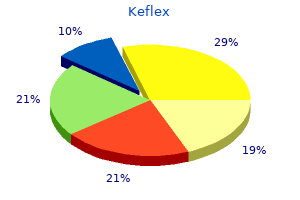
750 mg keflex order visa
Kenin A virus protection for windows xp keflex 500 mg discount otc, Levine J bacteria dies at what temperature keflex 500 mg order with mastercard, Spinner M: Parosteal lipoma: a report of two cases with related bone adjustments. Moghal N: Vascular and cartilaginous hamartoma (mesenchymoma) of the ribs in infancy. Ulloa-Patino P, Baeza-Flores E, Montalvo-Marin A, et al: Vascular and cartilaginous hamartoma of the thoracic wall. Eckardt A, Swennen G, Teltzrow T: Melanotic neuroectodermal tumor of infancy involving the mandible: 7-year follow-up after heminmandibulectomy and costochondral graft reconstruction. Franchi G, Sleilati F, Soupre V, et al: Melanotic neuroectodermal tumour of infancy involving the orbit and maxilla: surgical administration and follow-up technique. Hoshino S, Takahashi H, Shimura T, et al: Melanotic neuroectodermal tumor of infancy in the skull associated with excessive serum levels of catecholamine: case report. Melanotic neuroectodermal tumor of infancy (melanotic progonoma) involving the calvaria. Khoddami M, Squire J, Zielenska M, et al: Melanotic neuroectodermal tumor of infancy: a molecular genetic research. Nitta T, Endo T, Tsunoda A, et al: Melanotic neuroectodermal tumor of infancy: a molecular approach to diagnosis-case report. Shokry A, Briner J, Makek M: Malignant melanotic neuroectodermal tumor of infancy: a case report. Bonetti F, Pea M, Martignoni G, et al: the perivascular epithelioid cell and related lesions. Bonetti F, Martignoni G, Colato C, et al: Abdominopelvic sarcoma of perivascular epithelioid cells. Jundt G, Moll C, Nidecker A, et al: Primary leiomyosarcoma of bone: report of eight circumstances. Kawai T, Suzuki M, Mukai M, et al: Primary leiomyosarcoma of bone: an immunohistochemical and ultrastructural study. Lee E, Locker J, Nalesnik M, et al: the association of EpsteinBarr virus with clean muscle tumors occurring after organ transplantation. Gaffey M, Mills S, Askin F, et al: Clear cell tumor of the lung: a clinicopathologic, immunohistochemical, and ultrastructural research of eight instances. Lantuejoul S, Isaac S, Pinel N, et al: Clear cell tumor of the lung: an immunohistochemical and ultrastructural examine supporting a pericytic differentiation. Yamamoto H, Oda Y, Yao T, et al: Malignant perivascular epithelioid cell tumor of the colon: report of a case with molecular evaluation. A novel member of the household of lesions characterised by the presence of perivascular epithelioid cells. Takemori M, Nishimura R, Sugimura K, et al: Thoracic vertebral bone metastasis from uterine leiomyosarcoma. Lamovec J, Zidar A, Bracko M, et al: Primary bone sarcoma with rhabdomyosarcomatous part. Oda Y, Tsuneyoshi M, Hashimoto H, et al: Primary rhabdomyosarcoma of the iliac bone in an adult: a case mimicking fibrosarcoma. Llombart-Bosch A, Contesso G, Peydro-Olaya A: Histology, immunohistochemistry, and electron microscopy of small round cell tumors of bone. Mrad K, Sassi S, Smida M, et al: Osteosarcoma with rhabdomyosarcomatous component or so-called malignant mesenchymoma of bone. Van Dorpe J, Sciot R, Samson I, et al: Primary osteorhabdomyosarcoma (malignant mesenchymoma) of bone: a case report and evaluation of the literature. In 1913, Fischer17 described a peculiar tumor occurring predominantly in the tibia and infrequently in the fibula that was somewhat just like a more widespread odontogenic adamantinoma of the jaw bones. Despite histologic similarity, there has been no proof that these tumors-of the jaw bone and of the lengthy bone-have a similar histogenetic origin. Adamantinoma of long bones is extremely uncommon; there are roughly 300 welldocumented circumstances printed on the planet literature. The morphologic variety of lengthy bone adamantinomas also accounts for the continuing controversy relating to their histogenesis. Another attention-grabbing phenomenon is the association of these tumors with areas resembling osteofibrous dysplasia (ossifying fibroma of long bones). The first description of areas of rarefaction related to lengthy bone adamantinoma interpreted to be osteitis fibrosa was supplied in 1942 by Dockerty and Myerding. The first comprehensive description of long bone adamantinoma related to areas resembling fibrous dysplasia was provided in 1962 by Cohen et al. Therefore these authors were the first to postulate that, regardless of the histologic similarity to fibrous dysplasia, fibroosseous lesions associated with adamantinoma may symbolize a distinctive function associated to these tumors. The description of ossifying fibroma of long bone by Kempson38 in 1966 and the next redesignation of this lesion as osteofibrous dysplasia by Campanacci et al. Further studies of such composite lesions confirmed that the histologic and radiologic features of the fibroosseous components were more consistent with those of osteofibrous dysplasia. The second group is defined as differentiated adamantinoma and is characterized histologically by a predominance of an osteofibrous dysplasia�like pattern with a small, inconspicuous component of epithelial tumor components. On radiographs, differentiated adamantinomas are intracortical and sometimes multicentric lesions. The histogenesis of those peculiar neoplasms remains to be unclear, and varied ideas of their origin as derived from eccrine gland, synovial, vascular, or pluripotential mesenchyma have been postulated. In the outline in this chapter, basic and differentiated adamantinoma are mentioned individually. It has an total histologic similarity to the more frequent odontogenic adamantinoma of the jaw bones. Incidence and Location Classic adamantinoma is extremely rare; while its true incidence is unknown, it clearly accounts for less than 1% of all bone neoplasms. The youngest patient with traditional adamantinoma in our sequence was a 13-year-old boy who subsequently developed lung metastases. Synchronous involvement of the tibia and fibula by two unbiased foci or by contiguous foci is typical of adamantinoma. Clinical Symptoms Pain, bowing deformity of the tibia, or both of those signs are essentially the most frequent scientific findings, and these could also be current for many years before analysis. Radiographic Imaging Features of classic adamantinoma on radiographs are distinct and often diagnostic. In the tibia, involvement of the anterolateral cortex and the presence of a reasonable to extreme bowing deformity are typical. On radiographs, some lesions appear as destructive lytic defects involving two adjoining bones, the tibia and fibula. The synchronous involvement by separate foci within the tibia and fibula is much less frequent in this type of adamantinoma.
Keflex 750 mg discount amex
Hyckel P treatment for dogs with diarrhea imodium cheap 750 mg keflex with amex, Berndt A fever after antibiotics for sinus infection cheap keflex 500 mg free shipping, Schleier P, et al: Cherubism-new hypotheses on pathogenesis and therapeutic consequences. Ishinaga H, Otsu K, Mouri G, et al: Aggressive large cell reparative granuloma of the nasal cavity. Subasi M, Kapukaya A, Buyukbayram H, et al: Giant-cell reparative granuloma of the tibia. Vered M, Buchner A, Dayan D: Central large cell granuloma of the jawbones-new insights into molecular biology with medical implications on treatment approaches. Von Wowern N: Cherubism: a 36-year long-term follow-up of 2 generations in different households and review of the literature. Yoshida T, Sakamoto A, Tanaka K, et al: Alternative surgical remedy for giant-cell reparative granuloma within the metacarpal, using phenol and ethanol adjuvant remedy. This lesion was distinguished from the final category of unclassified round-cell sarcomas and lymphohematopoietic tumors based mostly on its clinicopathologic options. The consistent presence of a chromosomal abnormality- the reciprocal translocation of chromosomes eleven and 22, which entails bands q24 and q12 of the chromosomes, respectively-is a unique finding on this group of tumors. During the previous three many years, molecular research have elucidated the structure of the chromosomal breakpoint and have recognized the fusion companions of the chimeric gene. There can additionally be hope that the evolving molecular knowledge can information us to more effective focused therapies for these tumors. The emergence of molecular strategies as a major aid in the analysis of this group of malignancies should be particularly emphasized. The description of molecular features of this group of tumors is targeted on the character of translocations and their genes as potential biomarkers in classification and differential diagnosis. The interested reader is referred to Chapter 3, the place the biologic results of signature hybrid proteins on this group of tumors are described. These translocations drive the biology of tumors, are diagnostically particular, and will represent future therapeutic targets. Regardless of whether or not the breakpoint is within intron 7 or 8, the exon eight sequence is spliced out of the chimeric transcript. The second component in the commonest fusion variant is the human homolog of the murine Fli1 gene positioned on chromosome 11q24. The molecular downstream targets for chimeric proteins are nonetheless poorly understood. Large solid arrows indicate probably the most frequent locations of the breakpoints within introns, and the open arrows point out much less frequent variants. B, Functional domains of the two proteins involved within the translocation are depicted. In concert with extra cofactors, it acts as a transcription issue, resulting in aberrant activation of genetic targets that drive the pathogenesis of this tumor. C, Computer processed pseudocolored image of the karyogram of the case shown in A and B. However, when such a rearrangement occurs within the background of complicated chromosomal abnormalities, its standard cytogenetic identification could also be problematic. They appear to functionally substitute these extra widespread translocations in oncogenic transformation. Considering the similarities between these two fusion partners, it is very doubtless that each chimeras use comparable mechanisms to orchestrate an overlapping repertoire of goal genes. C, Functional domains of the two proteins involved in the translocations are depicted. It seems that the tendency for pluripotential differentiation is extra typically seen in lesions involving the thoracopulmonary area. Note that every of those genes has utterly distinct amino acid composition as properly as useful domains. It is essentially the most frequently occurring major sarcoma of bone after osteosarcoma and chondrosarcoma. With the appearance of molecular probes and immunohistochemical techniques, differentiation amongst these tumors hardly ever presents a diagnostic problem. The perpendicular "sunburst" type of periosteal new bone formation may be present however is less widespread compared with its occurrence in osteosarcoma. The lesion entails the medullary cavity and soft tissue extra extensively than is often documented Text continued on p. A, Plain radiograph of forearm of a 19-year-old girl with giant delicate tissue mass surrounding intramedullary tumor of proximal radial shaft. B and C, T1- and T2-weighted magnetic resonance pictures show in depth intramedullary tumor involving radial shaft and together with head of radius. Bulky gentle tissue mass developed by tumor permeating via cortex with out leaving gross evidence of destruction in plain movies. A and B, Lateral and anteroposterior radiographs of the femur in a 20-yearold girl show a harmful intramedullary tumor with lamellated periosteal reations; margins are sick defined. Image on left reveals intramedullary tumor and huge extraosseous mass in posterior muscle group. A, Plain radiograph of humerus of a 19-year-old man reveals massive area of diaphyseal destruction with permeative options. B, T1-weighted magnetic resonance picture of tumor shown in A reveals large extraosseous mass and intramedullary tumor. A, Anteroposterior radiograph reveals a damaging lytic lesion involving the distal fibula. B, Coronal magnetic resonance image of the lesion in A exhibits high sign intensity in an intramedullary tumor with extensive involvement of the parosteal gentle tissue. C, Radioisotope bone scan of the identical lesion exhibits elevated uptake in the best distal femur. A, A-P radiograph exhibiting a permeative lesion of femoral shaft with extension into soft tissue. Note radiating periosteal new bone formation and concave cortical defect (saucerization). E, Gross photograph of the identical case proven in C and D exhibits intramedullary mass permeating overlying thickened cortex with subperiosteal mass. A, Permeative lesion of proximal femoral shaft associated with cortical thickening. Magnetic resonance imaging and computed tomography allow extra accurate preoperative evaluation of the lesion than plain radiographs. They usually reveal intramedullary lesions that contain giant segments of the intramedullary cavity and extend beyond the area shown to be concerned on plain radiographs. This type of radiographic appearance is typically seen in predominantly subperiosteal lesions.
Buy keflex 750 mg cheap
Moreover antibiotic medications keflex 500 mg order with mastercard, not all tumor cells may develop all properties listed; some cell clones could subspecialize antibiotics mirena 250 mg keflex. B, Summary of molecular modifications related to tumor cell interplay with numerous stromal components important for invasive development and metastasis. Integrins, a superfamily of transmembrane receptors that modulate cell-tocell and cell-to-matrix interactions by binding with a big selection of ligands, play a key role in skeletal metastases on many levels, matrix and blood vessel invasion, osteoclast signaling, neovascularization, and colonization of bone. Thus the interaction of tumor cells with extracellular matrix seems to be one of the main mechanisms of the metastatic cascade. The basement membrane is a sheetlike structure that separates epithelial and endothelial cells from the interstitial matrix. This large complicated molecule has three quick arms containing collagen binding websites and a single lengthy arm with a heparan binding website. The central region of the molecule, where all four arms are connected in a cross-shaped structure, incorporates the laminin receptor binding web site for cells. A, Both systemic factors and locally performing factors induce the formation and activity of osteoclasts. Accessory cells such as T cells can produce cytokines that can inhibit the formation of osteoclasts, similar to interleukin-4, interleukin-18 and interferon-. B, Both systemic elements and domestically acting components can enhance the proliferation and differentiation of osteoblasts. In addition, bone matrix is a significant supply of development factors, which might improve the proliferation and differentiation of osteoblasts. Fibronectin is one of the main noncollagenous parts of the interstitial matrix. To invade the intercellular matrix and to penetrate the vascular channels, the tumor cells need to observe three general steps: 1. They should maintain some degree of their attachment to the matrix elements (mainly laminin and fibronectin). Degradation of the matrix components should proceed via the action of proteolytic enzymes. Tumor cells must continue to migrate into the degraded space of the extracellular matrix. Paradoxically, it has been proven that tumor cells exhibiting increased ranges of laminin and fibronectin floor receptors have a higher metastatic potential compared with those that have minimal ranges of these receptors. Thus to develop metastatic foci, the tumor cells 1220 19 Metastatic Tumors in Bone should maintain a point of adherence to the intercellular matrix elements. In basic, some insufficiency of cell adhesion and junction methods may be documented in most cancers. Tumor cells that invade vessels have a higher capacity to degrade the stromal matrix than different tumor cells. The enzymatic activity of tumor cells plays an important function in the improvement of metastases. The involvement of several totally different proteases, together with urokinase, plasminogen activator, cathepsins B and D, and varied metalloproteases produced instantly by tumor cells, play an necessary function in the invasive progress and improvement of metastasis. Therefore their roles in invasive development and metastasis rely not solely on the level of the enzyme, but in addition on the presence of adequate amounts of their activators and inhibitors. The exercise of this enzyme may be correlated with the metastatic potential of several experimental and human tumors. Overall, an upregulation of multiple proteolytic enzymes of the so-called plasminogen cascade have been documented in malignant tumor cells and have been linked to their invasive and metastatic potential. The capability to induce vascular growth is another issue that secures the survival of an enlarging tumor mass. The capacity of tumor cells to induce proliferation of vascular cells through a variety of growth elements such as endothelial progress factor and fibroblastic progress factor has just lately been extensively studied. The tumor cells that invade the vessels and circulate within the lymph or blood interact with mobile and humoral parts of the environment. The interplay of tumor cells with platelets and other blood clotting components, both circulating and cell fixed, is a vital component within the promotion of tumor cell thrombosis of peripheral sinusoids and progress of metastatic foci. Moreover, the tumor cells of a metastatic focus should retain their stromal destructive activities and interact with different cells of the new setting to survive and type clinically detectable nodules. Osteolysis is driven by osteoclastic resorption of bone, the control of which comes beneath quite lots of influences. Carcinomas of the prostate and breast, as properly as neuroendocrine tumors, can induce predominately blastic or sclerotic metastases. These elements induce bone formation by inducing osteoclast apoptosis whereas stimulating osteoblast differentiation and proliferation. In addition, the vertebral plexus of veins is valveless, and the retrograde venous strain is commonly increased within the abdominal and chest regions. This allows a retrograde flow of blood to bypass the caval system and to reach the bones of the vertebral column. Thus the biologic circumstances of bone tissue must even be important factors in promoting the expansion of tumor cells that reach the marrow via the venous and arterial blood community. Autopsy reports of large collection of sufferers have proven that, with gross examination and restricted sampling, 19 Metastatic Tumors in Bone 1221 skeletal metastases could be documented in 30% of patients who died of carcinoma, with explicit carcinomas similar to breast or prostate cancer current in nearly 85% of patients autopsied. This is in line with observations by Jaffe, who acknowledged that if extensive skeletal sampling were to be performed, metastases could be documented in 70% of sufferers who died of carcinoma. Therefore they might not detect metastatic tumors which are primarily related to bone destruction and minimal or no osteoblastic activity. Cortical disruption, extension into delicate tissue, and periosteal new bone formation could be current. Often, focal sclerosis is seen inside lytic lesions, and due to this fact many skeletal metastases produce a combination of lytic and blastic appearances. In common, a lytic versus blastic radiographic appearance of a metastatic tumor in bone results from a prevalent bone resorptive (destructive) or stimulating (osteoblastic) activity of the tumor. Consequently, such bones because the vertebrae, pelvis, ribs, cranium, sternum, proximal femur, and humerus are most frequently involved. These websites correspond to areas that include hematopoietic marrow, which has a rich sinusoidal vascular community. This feature and the presence of venous plexus connected with belly and thoracic organs may promote metastasis in these areas. Metastases that predominantly involve fatty marrow distal to elbow and knee joints and the mandible are unusual in adults. In some sufferers, it can be a presenting signal of a clinically silent main tumor that, most often, is situated within the thoracic or belly organs. Although it could possibly occur in many frequent cancers, carcinoma of the lung is essentially the most frequent malignancy in which so-called acral metastases happen. In common, acral metastases are most often seen in the small bones of the toes in extremely aggressive malignant neoplasms of visceral and thoracic organs.
Purchase 750 mg keflex free shipping
Cystic Angiomatosis the term cystic angiomatosis is utilized to extraordinarily rare situations of disseminated multifocal hemangiomas within the skeleton antibiotics for uti not sulfa generic 500 mg keflex with mastercard. The sites of extraskeletal involvement include delicate tissue antimicrobial bed sheets 750 mg keflex fast delivery, lung, liver, and significantly spleen. The condition can be asymptomatic and is often detected incidentally in radiographs obtained for different reasons. Radiolucent lesions with a soap-bubble or honeycomb appearance are current in the cranium, backbone, ribs, and pelvis. In rare situations, the dysfunction is manifested on radiographs as disseminated osteoblastic lesions that mimic osteoblastic metastases. A and B, Lymphangiomatosis of the rib displaying cortical bone eroded by medullary lymphangiomatous course of composed of dilated, endothelial-lined channels. C and D, Dilated lymphatic channels of various sizes forming a spongy architecture inside the medullary bone. A, Radiograph of fibula exhibits area of cortical expansion with coarse trabeculation. B, Gross photograph of bivalved resection segment of fibular shaft shows localized space of honeycomb appearance positioned eccentrically in cortex. C and D, Low and intermediate power photomicrographs of lymphangioma of bone show dilated, endothelial-lined channels with delicate walls. A, Chest radiograph of a 7-year-old boy with lymphangiomatosis of a number of proper ribs and right-sided chylous effusion. B, Coarse trabeculation of multiple ribs on proper side was produced by lymphangiomatosis. C, Computed tomogram of chest shows chylous effusion and rarefaction of posterior portion of rib. On radiographs, lytic lesions may be seen to contain some residual lamellar bone and foci of reactive woven bone. Blastic lesions comprise prominent, thickened bone trabeculae of mature lamellar bone with areas of woven bone rimmed by osteoblasts. Evaluation of serial radiographs on this dysfunction has shown that growing osteosclerosis can be an age-related phenomenon in angiomatous foci which may be initially lytic. It may be that the lesions spontaneously heal or regress and that this process is accompanied by sclerosis. Rare cases of angiosarcoma arising from skeletal angiomatosis have been described. Usually, it includes the shoulder and hip areas and begins in the trunk bones, and it could individually involve a number of bones in the same area. In the appendicular skeleton, the proximal components of the extremity bones-the proximal humerus and femur-are typically involved. They are indistinguishable from strange, nonaggressive angiomatoses and represent a complex Text continued on p. B, Multiple lucent areas with coarse trabeculation involving patella, proximal tibial metaphysis and epiphysis, and proximal fibular shaft in teenaged patient. A, Computed tomogram of pelvis and sacrum of 60-year-old man with sclerosing variant of cystic angiomatosis. B, Radionuclide bone scan shows elevated uptake in parts of pelvis exhibiting sclerotic lesions in A and in left second and fifth ribs. A, Sclerotic variant of cystic angiomatosis involving each left and right pelvic bones. B, Sclerotic lesions mimicking osteoblastic metastases adjacent to sacroiliac joint and iliac crest. C, Low energy photomicrograph shows vascular lesion overgrown by well-developed thickened trabeculae of bone (C, �25) (C, hematoxylin-eosin. Note multifocal sclerotic lesions mimicking osteoblastic metastasis adjoining to sacroiliac joints. A, Low power photomicrograph shows dilated, thin-walled vessels in marrow spaces of cancellous bone with delicate trabeculae of reactive woven bone surrounding angiomatosis parts. B, Medium energy photomicrograph exhibits thin-walled vascular channels in marrow spaces bordered by thickened mature bone trabeculae (A and B, �50; hematoxylin-eosin). Note vascular channels in marrow areas surrounded by thick trabeculae of lamellar bone resembling cortical bone (�50). B, Medium energy photomicrograph of A reveals osteoclastic resorption cavity (right) and embedded vascular structures (�100). The energetic lesions are purely vascular, and the osteoclastic resorptive exercise could be seen near the advancing edge. Stabilization of the lesion is associated with an elevated amount of intervening connective tissue. In some cases, extensive involvement of the chest wall (ribs) is related to pleural effusion and extreme pulmonary compromise, which might result in a deadly consequence. Because the clinical course of massive osteolysis is variable and unpredictable, the selection of a specific methodology of remedy is fraught with uncertainty. In reality, any histologically benign angiomatosis of bone or gentle tissue may sometimes behave clinically as a slowly progressive dysfunction that eventually compromises the perform of the affected organ. In such instances, main resection procedures including amputation could additionally be required in order to control the method. Surgical resection, embolization, radiation therapy, steroids, and more lately, interferon alfa have all been used in instances showing progression. They are classified similarly to vascular neoplasms as of arterial, venous, or capillary in nature. They are predominantly gentle tissue lesions and their involvement of the skeleton is quite secondary. These conditions are greatest recognized in correlation with clinical presentation and radiographic imaging knowledge. Glomus Tumor the glomus body is a type of arteriovenous anastomosis that performs a task in thermal regulation. It is distributed throughout the body in the reticular dermis however is most frequently seen in the subungual region. The normal glomus physique consists of an afferent arteriole that branches into two to 4 connecting arterioles. The arterioles have thick segments surrounded by concentric layers of modified perivascular easy muscle cells. These segments are referred to as Sucquet-Hoyer canals and include 932 13 Vascular Lesions single most common site is the subungual region of the fingers. They can also occur within the tip of the coccyx, where they arise from the glomus coccygeum. Exceptional cases of glomus tumor arising in other bones such as the proximal phalanx, tibia, ulna, and pelvis have also been reported. Clinical Symptoms Glomus tumors are extremely painful, and symptoms are often out of proportion with the dimensions of the tumor. Paroxysms of ache in the affected space may be triggered by changes of temperature or even by minor mechanical disturbance of the affected area. Intraosseous lesions that cause these signs increase clinical suspicion of osteoid osteoma.
Buy keflex 750 mg free shipping
Subsequent expertise has proven that the use of numeric histologic grading lacks predictive worth infection nosocomial purchase keflex 750 mg with visa. The classic microscopic sample of large cell tumor is frequently modified by a secondary reactive proliferation of fibrohistiocytic tissue antibiotic for dogs discount 750 mg keflex amex, hemorrhage, necrosis, and aneurysmal bone cyst formation. Reactive fibrous tissue with a outstanding storiform sample and xanthogranulomatous reaction can be documented, at least focally, in practically all appropriately sampled lesions. A, Anteroposterior radiograph of big cell tumor in skeletally immature patient exhibits no involvement of epiphysis. B, Lateral view of tumor proven in A shows no extension to secondary growth heart. C, Lateral radiograph of distal femur of a 27-year-old lady with ache in lower thigh. D, T1-weighted magnetic resonance picture exhibits that giant cell tumor seen in C is limited to shaft. E, Bisected distal radial resection specimen showing an expansile multilocular big cell tumor composed of dark and light tan tissue with focal yellowish areas. A, Anteroposterior radiograph displaying an expansile lytic lesion of the proximal fibula with fine trabecular sample. B, Bisected proximal fibula resection specimen displaying an expansile giant cell tumor with multilocular cystic structure. A, Bisected distal femoral resection specimen reveals mild and dark tan large cell tumor with marbling fibrous tissue. Mild expansion of the bone contour with intact cortex and sharp demarcation of the tumor from the encompassing medullary bone is present. B, Expansile giant cell tumor of distal femur composed of sunshine and darkish tan tissue with central fibronecrotic areas. C, Giant cell tumor of tibia extending to the articular plate and expanding the bone contour laterally. D, A large expansile giant cell tumor of the proximal humerus composed of light and dark tan tissue. Large areas of yellowish necrotic tissue within the central portion of the lesion are present. B, Posterior aspect of specimen proven in A shows destroyed cortex and growth of tumor into popliteal fossa. C, Bisected distal finish of radius with well-demarcated eccentric tumor mass expanding bone contour. A, Classic microscopic sample of big cell tumor with oval or plump spindle mononuclear cells uniformly interspersed with multinucleated big cells. D, Higher magnification of C exhibits mononuclear histiocytic cells and multinucleated giant cells with hemosiderin deposits. B, Higher energy view of A exhibits multinucleated large cells and oval or plump mononuclear cells. C, Classic microscopic features of large cell tumor composed of mononuclear histiocytic cells with evenly distributed multinucleated big cells. B, Higher power view of A exhibits multinucleated large cells with greater than a hundred nuclei and dense eosinophilic cytoplasm. C, Conventional giant cell tumor with spindling of mononuclear cells and scattered multinucleated big cells with ragged contours of their cytoplasm. D, Higher magnification of C showing multinucleated giant cells with massive irregular cytoplasm. A-H, Spectrum of multinucleated large cells incessantly seen in big cell tumors ranging in size of their cytoplasm and the number of nuclei from a couple of to several hundred. A, Xanthogranulomatous response in large cell tumor obliterating its classical cytoarchitectural features. Ill-defined bands of spindle-cell proliferations interspaced with histiocytic infiltrate are present. D, Higher magnification of C exhibiting blended fibrous and histiocytic infiltrate with occasional atypical cells. A-D, Giant cell tumor exhibiting enlargement and nuclear atypia of the mononuclear histiocytic cells. Occasionally, fibrohistiocytic reaction massively replaces the underlying tumor in order that it mimics lesions corresponding to nonossifying fibroma or benign fibrous histiocytoma. In such situations, the analysis of radiographic information and additional sampling of the tumor are normally enough to document the existence of an underlying big cell tumor. Prominent focal reactive bone sometimes may be correlated with the presence of small cortical infractions. This peculiar capability of big cell tumor to induce reactive peripheral ossification is maintained in recurrences in gentle tissues, in pulmonary implants, and even in the transplanted fragments of tumor tissue to athymic nude mice. Hemorrhage, necrosis, or both normally end result from fracture or mechanical compression. Old and fresh hemorrhage, in addition to necrosis, may be present with none obvious trigger and can be quite intensive. For unknown causes, the mononuclear stromal cells normally develop the recognizable options of necrosis first. It is common to observe well-preserved big cells in a very necrotic stroma. The focal nature of this necrosis-related atypia in an otherwise conventional giant cell tumor and the absence of atypical mitoses are useful in avoiding a misdiagnosis of malignant change. Microscopic foci of aneurysmal bone cyst may be regularly documented if acceptable samples can be found. A large cell tumor is reported to be an underlying situation in 10% of secondary aneurysmal bone cysts. On the opposite hand, solid areas containing quite a few multinucleated big cells in a spindle-cell stroma are frequently present in an aneurysmal bone cyst and can be readily misinterpreted as an underlying large cell tumor. This discovering must be interpreted only as regards to appropriate radiographic options and medical setting to avoid misdiagnosis of large cell tumor. The overall similarity of these proliferations to nodular fasciitis is helpful to distinguish such benign reactive processes from sarcomatoid transformation. Multinucleated big cells are much like osteoclasts however often include many extra nuclei. Characteristically, the nuclei of mononuclear histiocytoid cells are similar to nuclei of giant osteoclast-like cells. The cytologic options of large cell tumor of bone are sometimes obscured by secondary changes, corresponding to proliferation of fibrous tissue accompanied by foamy histiocytes. In such cases, correlation of cytologic findings with clinical and radiologic knowledge might help to set up the right diagnosis. Differential Diagnosis Giant cell tumor must be differentiated from large cell reparative granuloma and different reactive big cell� containing lesions, such because the brown tumor of hyperparathyroidism.
Keflex 250 mg lowest price
D bacterial cell diagram order keflex 500 mg without prescription, Higher magnification of C exhibiting mobile pleomorphism and nuclear atypia of tumor cells antibiotics for uti can you drink alcohol discount 750 mg keflex. Inset, Higher magnification of two highly atypical tumor cells with dense oval cytoplasm. After authentic remedy, the tumor showed a quantity of native recurrences with invasion and satellite tv for pc nodules within the adjoining gentle tissue over a period of practically a decade. The perturbation of Hedgehog and Wnt signaling pathways is considered to disrupt the pattern of enchondral ossification in the development plate, resulting in faulty formation of the bony collar. Definition Solitary osteochondroma is a developmental anomaly of bone that ends in the formation of an exophytic outgrowth on the floor of bone. Incidence and Location Solitary osteochondroma is the most common benign tumorlike lesion, accounting for roughly 35% of benign bone tumors and 10% of all bone tumors in any major series. Osteochondromas have a definite male intercourse predominance, and the male-to-female intercourse ratio is 1. Osteochondromas have a predilection for the appendicular skeleton and most frequently happen on the floor of metaphyseal parts of major lengthy tubular bones. The knee area is most frequently affected, with roughly 35% of instances occurring in this web site. Osteochondromas also frequently happen in the flat bones and often contain the ilium and scapula. They are uncommon in small tubular bones of the arms and feet, in the ribs, and within the vertebral column. In rare cases a fracture, usually on the base of the stalk, could be a presenting symptom. A bursa may be current over the cap, which can turn into inflamed or accumulate synovial fluid or free bodies, thereby producing symptoms. The stalk is regularly slender, and its extremity is covered by a lobulated cartilaginous cap which will comprise calcifications. The stalk can measure a quantity of centimeters along its long axis, however the cartilage cap is often thin (2 to 3 mm). A lengthy bone harboring a solitary osteochondroma can present slight localized distortion of the contour at its base, however the overall modeling deformity, clubbing of the bone finish, and development disturbance typical of multiple hereditary exostoses are absent. These scans are additionally useful in establishing the continuity of the lesion with the adjacent cortex and underlying spongiosa. An overlying bursa (exostosis bursata) may be present external to the cartilaginous cap. The physique and stalk of an osteochondroma additionally may present irregular calcific densities that represent unresorbed calcified cartilage. The cut floor reveals the hyaline composition of the cap, which often measures from 2 mm to 1 cm in thickness. This reflects the endochondral ossification sequences present Text continued on p. A, Pedunculated osteochondroma of the proximal tibial metaphysis in a skeletally immature particular person. B, Sessile osteochondroma of the proximal fibula and in a skeletally immature individual. Note the continuity between the inner house of osteochondroma and the underlying medullary cavity of the concerned bone. C, Pedunculated osteochondroma of the distal femoral metaphysis in a skeletally immature individual. D, Pedunculated osteochondroma of the distal femoral metaphysis with synovial chondromatosis in bursa overlying the cartilage cap. D, Anteroposterior radiograph displaying a big broad-based osteochondroma arising from the neck of the left femur. E, Lateral radiograph showing a big broad-based osteochondroma arising from the posterior facet of the proximal tibia. Inset, T1-weighted axial magnetic resonance picture exhibiting a big osteochodromatous mass arising from the posterior aspect of the tibial surface. A and B, Surface and bisected broad-based osteochondroma arising from the proximal fibular metaphysis. C and D, Surface and bisected peduculated osteochondroma arising from the proximal femoral metaphysis. E and F, Bisected broad-based osteochondroma with a corresponding specimen radiograph arising from the proximal fibular metaphysis. E, Bisected resection specimen of the case in C and D exhibiting an irregular thick cartilage cap masking the floor of this osteochondroma. A-D, External surface and serial sections of a large broad-based osteochondroma arising from the iliac floor. A and B, Cut floor and macrophotograph of histologic sections present relationship of cartilage cap to underlying cancellous bone in sessile osteochondroma. C and D, Cut surface and whole-mount macrophotograph of slender pedicled osteochondroma present cartilage cap at surface. In adults the cartilage cap could also be extraordinarily thin or absent because endochondral ossification and progress of the osteochondroma cease at skeletal maturation or shortly thereafter. Deep to the cap, the central portion of the spongy bone may comprise irregular islands of calcified cartilage that can be seen grossly as gritty, opaque white deposits. The spongiosum of the stalk is continuous with the cancellous bone of the underlying medullary cavity and is bordered by compact cortical bone that flares from the adjacent cortex. The periphery is roofed by a fibrous perichondrium that blends imperceptibly with the outer layer of hyaline cartilage. As cell replication diminishes in the cartilage cap, its thickness diminishes, and it may entirely disappear in older adults. The spongy bone beneath the cap may include one or more irregular masses of necrotic calcified chondroid, which represent unresorbed parts of the calcified zone of the cap. The cartilage cap may show foci of increased cellularity, and individual atypical and even multinucleated chondrocytes could also be current. The bursa that will form on the periphery of some osteochondromas is often intimately hooked up to the perichondrium of the cap. In some instances, chondroid metaplasia on this bursal synovial lining may be associated with quite a few cartilaginous unfastened bodies in the bursa. If calcified, these metaplastic chondroid synovial nodules can simulate secondary chondrosarcoma on radiographs. Differential Diagnosis crucial lesion to distinguish from osteochondroma is parosteal osteosarcoma. This floor malignancy of bone is characteristically more radiodense than an osteochondroma, especially at the base and heart of the bony excrescence. The cartilage cap sometimes found on the periphery of a parosteal osteosarcoma differs from that of an osteochondroma because it contains cytologically malignant cartilage cells with out evidence of enchondral ossification at the interface with the underlying bony and spindle-cell components. Unless a fracture has occurred within the stalk of an osteocartilaginous exostosis, the cancellous bone consists of mature lamellar trabeculae with fatty marrow. Continuity with the marrow cavity of the underlying bone may be found constantly in osteochondromas, and an intact cortex is usually current beneath a parosteal osteosarcoma. Juxtacortical myositis ossificans and other posttraumatic surface lesions (florid reactive periostitis, subungual exostosis, bizarre parosteal osteocartilaginous proliferation) may additionally be confused with an osteocartilaginous exostosis.
Generic 750 mg keflex fast delivery
Only after these elements have been thought of should the histologic options be studied and a diagnostic conclusion reached in the context of all obtainable scientific and radiographic information antibiotic youtube cheap 750 mg keflex fast delivery. Omission of any of those steps or taking "shortcuts" to the diagnosis of a cartilage tumor is fraught with danger antibiotics for uti breastfeeding discount keflex 250 mg visa. Serious errors have been made in overdiagnosis of incidentally discovered enchondromas, in addition to underestimation of medullary cartilage tumors, which, on reflection, confirmed indisputable radiologic options of aggressiveness. Central cartilage tumors of the ribs, sternum, and pelvis are unlikely to be benign, particularly if they exceed 4 cm in dimension. The restricted experience with these lesions signifies that male sufferers are probably more incessantly affected than are female patients. The acral skeleton with the involvement of the quick tubular bones of the hands is the second most frequently affected site. Rare examples of periosteal chondroma have been described in the backbone, clavicle, ribs, and toes. Because periosteal chondromas are frequently found near tendon insertion sites and disturb their perform, pain and local discomfort on activity could be initial signs. In a typical case, the cartilaginous nature of juxtacortical chondroma is straightforward to recognize on plain radiographs. The affected space can also show minimal evidence of a zone of subcortical sclerosis beneath the lesion. The cortex beneath is eroded and usually smoothly excavated, however generally it has a scalloped border. The lesion is clearly separated from the medullary cavity by a rim of sclerotic cortical bone. On average, juxtacortical chondromas are more cellular than are enchondromas of lengthy bones and can present gentle nuclear atypia or binucleated chondrocytes. A and B, Lateral and oblique plain radiographs of unusually large periosteal chondroma of proximal humerus in younger man. A, Plain radiograph reveals concave cortical erosion close to insertion of biceps muscle on lateral side of humeral shaft. Note sharply defined borders of cortical erosion and intact cortex beneath lesion. D, Gross photograph of periosteal chondroma of humerus reveals chondroid tumor beneath periosteum, eroding underlying cortex. A, T1-weighted sagittal magnetic resonance picture shows periosteal lesion rising on the popliteal surface of the femur. B, Whole-mount photomicrograph exhibiting a fibrous capsule similar to elevated periosteum masking the surface of the lesion and the partially eroded cortical bone beneath the lesion. C and D, Low energy photomicrographs exhibiting increased cellularity and clustered development pattern of chondrocytes (C and D, �50). A, Gross photograph of periosteal chondroma exhibits chondroid tumor beneath periosteum eroding underlying cortex. B, Composite whole-mount photomicrograph of periosteal chondroma displaying a cap-shaped lesion growing beneath the periosteum and eroding the underlying cortex. C and D, Low power photomicrographs showing base of periosteal chondroma with interface between lesion and underlying cortex. Note scalloping of underlying cortical bone, but there is generally a sharp demarcation between periosteal chondroma and cortex with no feature of infiltrative aggressive development sample (C and D, �40). A and B, Low power photomicrographs of the lateral aspect of periosteal chondroma displaying elevations of periosteum and erosion of the underlying cortex. C and D, Low energy photomicrographs of the base of periosteal chondroma showing erosion of underlying cortex. A-D, Low energy photomicrographs showing satellite nodules in periosteal chondromas. The satellite tv for pc nodules are usually present within the superficial and lateral elements of the lesion. A, Lobulated cartilaginous tumor bordered by extensive subperiosteal lamellar bone. Apparent satellite nodules at periphery (arrows) characterize tangentially minimize marginal irregularities. B, Nodular marginal extension of periosteal chondroma with central calcification and endochondral ossification. Inset, High cellularity and delicate atypia of chondrocytes (A, �20; B, �60; inset, �200). Special Techniques Similar to enchondromas, periosteal chondromas expresses a full roster of markers characteristic of cartilage lineage differentiation. In such a way, these lesions are immunohistochemically indistinguishable from ordinary enchondromas. Several distinct clonal chromosomal abnormalities have been described in periosteal chondromas; they include rearrangements of 2q37, 4q21-25, 11q13-15, and 12q13. Differential Diagnosis Juxtacortical chondrosarcoma can usually be distinguished with ease by the massive size of the lesion (>5 cm) and the absence of radiologic evidence of solid periosteal new bone buttressing on the margins. Microscopically, the diploma of cellularity, variation in dimension and shape of nuclei, and frequent multinucleate chondrocytes are the premise for differentiating these two tumors. Periosteal osteosarcoma, a predominantly cartilaginous form of floor osteosarcoma, can be acknowledged by the feathery perpendicular calcific striae seen on radiographs. Periosteal osteosarcoma is diagnosed from the presence of sheets of primitive mesenchymal cells between the cartilage lobules, with tumor osteoid and bone deposition between the cells. Treatment and Behavior Because periosteal chondromas might show radiologic overlap with juxtacortical chondrosarcoma, the preferable mode of remedy is extensive native excision that includes the underlying cortex. In some cases periosteal chondroma might trigger development disturbance of the affected bone. In such circumstances the presence of the lesion is related to shortening of the bone. The term dyschondroplasia was introduced by Ollier in his description of the entity in 1900. The lesions have a tendency to be metaphyseal and are generally eccentrically positioned, with predominant unilateral involvement of the appendicular skeleton. The medical manifestations sometimes seem throughout childhood, and the extent of skeletal involvement is variable. The largest sequence reported from the Rizzoli Institute consisted of 51 circumstances of a quantity of enchondromas presumably representing enchondromatosis in contrast with 334 solitary enchondromas encountered in the identical interval. On the other end of the spectrum are the cases with large involvement of multiple bones and severe deformities. The most frequent presentation is a tumor affecting one extremity, however bilateral involvement is often current. Even in instances with diffuse involvement of multiple bones, the illness tends to predominate on one facet of the physique. The bones most often affected after the hand are the brief tubular bones of the ft, femur, and humerus and the bones of the forearm. The femur is the most regularly involved long tubular bone, followed by the tibia and humerus. Clinical Symptoms In common the extra diffuse and severe the skeletal involvement, the earlier in life symptoms seem.
Buy keflex 250 mg visa
The trabecular pattern of tumor bone sometimes focally mimics the looks of benign reactive woven bone or that of benign osteoblastic tumors antibiotics japan over counter keflex 750 mg. The main difference is within the cells that fill the intertrabecular areas virus yahoo email cheap keflex 250 mg fast delivery, which in osteosarcoma exhibit atypical, sarcomatous features. The infiltrating pattern within such areas, with destruction or engulfment of preexisting nontumor bone, facilitates the prognosis of a malignant process. These tumors may have a deceptively benign look and may be mistaken for osteoblastomas of the standard or aggressive type (for a extra detailed description see Chapter four and the part on osteosarcoma with distinctive microscopic features on this chapter). The cartilaginous differentiation in osteosarcoma may be very heterogeneous and represents all levels of cartilaginous matrix formation. The earliest levels symbolize the formation of a free basophilic intercellular substance with cells lying in lacunar spaces. Mineralization of the matrix produces bluish granular or linear deposits of calcium. Other patterns of cartilage differentiation parallel those seen in chondrosarcoma and vary from areas just like those seen in high-grade chondrosarcoma through myxoid change to well-differentiated areas with a deceptively benign look. Often a full spectrum of cartilage differentiation representing completely different degrees of matrix maturation and cellular atypia is seen in one tumor. Even in a predominantly chondroid tumor, a minimum of focal direct osteoid manufacturing by sarcomatous tumor cells is required for the recognition of its osteosarcomatous nature. Fibroblastic differentiation in osteosarcoma is characterised by the presence of a predominant spindle-cell part just like that seen in fibrosarcoma. The production of tumor bone by fibroblast-like cells discloses the bone-forming nature of these predominantly spindle-cell lesions and helps differentiate them from different spindle-cell neoplasms of bone. Such tumors, if present in an acceptable medical setting, are still finest categorised as osteosarcoma. Furthermore, these almost purely fibroblastic lesions occasionally can produce very closely ossified metastases. Low-grade fibroblastic osteosarcomas (intramedullary and surface) Text continued on p. B, Higher magnification of A showing lacelike sample of osteoid deposition and courts of anaplastic tumor cells. D, Higher magnification of C displaying interconnected seams of osteoid encircling tumor cells. A, Nearly stable sheetlike osteoid encircling small clusters of tumor cells with lacuna areas. C and D, Variants of seamlike osteoid deposition encircling small clusters of tumor cells and sometimes particular person tumor cells. B, Higher magnification of A showing loosely organized epithelioid tumor cells with oval densely eosinophilic cytoplasm. D, Higher magnification of C exhibiting early seams of unmineralized osteoid among tumor cells. B, Interconnected coarse deposits of osteoid encircling and separating clusters of tumor cells. D, Well developed interconnected irregular trabecular pattern produced by tumor osteoid. Note entrapment of individual tumor cells residing inside lacunar areas of osteoid. C, Conspicuous, well mineralized, osteoid deposition sample separating and encircling bigger clusters of loosely arranged tumor cells. D, Higher magnification of C exhibiting well mineralized osteoid and loosely arranged anaplastic tumor cells. B, Higher magnification of A displaying a less developed area of osteoid deposition with sparse seams of matrix deposition. B, Higher power of A exhibiting irregular stable areas of osteoid with entrapped tumor cells. C, Irregular stable areas of osteoid separating massive clusters of epithelioid tumor cells. Note dense eosinophilic cytoplasm responsible for epithelioid look of tumor cells. D, Higher energy of C showing irregular areas of osteoid with entrapped tumor cells. A and B, Infiltrative development pattern of high-grade osteosarcoma permeating intratrabecular marrow spaces. C, Higher energy of B displaying permeation of intratrabecular marrow areas by an osteosarcoma. D, Variegated osteoid deposition and mineralization patterns in osteosarcoma permeating marrow areas. A, Hemangiopericytoma-like sample of tumor progress bordering endothelial-lined areas. B, Higher magnification of A showing prominent vascular areas and sheetlike osteoid deposition. C, Hemangiopericytoma-like sample with outstanding vasculature and sheetlike osteoid deposition. D, Higher magnification of C displaying prominent engorged vessels and osteoid deposition by tumor cells. B, Higher magnification of A showing gradual transition between hypercellular sarcomatoid element and areas of more solid matrix deposition. D, Higher energy of C exhibiting a gradual transition between strong areas of anaplastic tumor cells and the cartilage matrix. A and B, Low and intermediate energy views of osteoid deposition by anaplastic tumor cells. C and D, Low and intermediate energy views of cartilaginous differentiation in the same tumor as shown in A and B. Occasionally a highly anaplastic tumor, sometimes located within the metaphysis of a long bone, exhibits no osteoid manufacturing but is in any other case cytologically much like an osteosarcoma. These lesions, previously categorized as undifferentiated (osteolytic) osteosarcoma, are actually designated as high-grade, pleomorphic, malignant fibrous histiocytoma of bone. The distinction is an academic one because both tumors are treated with similar chemotherapeutic regimens. By conference, a four-tier system has been used for histologic grading of typical osteosarcoma. Moreover, separation of conventional osteosarcoma into 4 grades relies on subjective analysis of nuclear atypia and appears to have minimal scientific value. The current trend is to separate typical osteosarcoma into two main categories: low and high grade.
