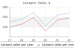
Cheap lexapro 10mg free shipping
This association explains why midline sections of the uterus bleed relatively less than lateral ones depression symptoms in young adults generic lexapro 10mg fast delivery. The tubal department courses laterally in the mesosalpinx near mood disorder support group lexapro 10 mg generic mastercard the uterine tube, which it supplies via a sequence of branches. The ovarian branch passes into the mesovarium, where it types a broad anastomosis with the ovarian artery proper from the stomach aorta. The anastomotic channel provides the ovary and offers a 16 Anatomy of the anterior belly wall, uterus, and pelvic organs sequence of tubal branches via the mesosalpinx that anastomose with the tubal department of the uterine artery. Both the uterine and ovarian arteries bear marked enlargement throughout pregnancy, the latter vessel bringing blood to the uterus through its broad anastomosis with the ovarian department of the uterine artery. The tortuous association of the ascending branch of the uterine artery allows the vessel to elongate and accommodate to the expansion of the uterus during being pregnant. The unspiraled descending branches are felt easily along the lateral side of the cervix at vaginal examination on account of their increased measurement during pregnancy. A huge ipsilateral and contralateral arterial anastomotic community forms throughout the uterus. Near the lateral pelvic wall, the uterine veins unite to type often two trunks that drain into the interior iliac vein. The main portion of venous blood from the uterus is drained by the use of the uterine veins. On the best, the ovarian plexus drains superiorly through the suspensory ligament of the ovary, crosses the best ureter obliquely, after which empties into the inferior vena cava. The ovarian veins on either side dilate to monumental measurement throughout pregnancy and are potential routes for thrombus formation. Dissection throughout cesarean section must remain close to the midline to avoid excessive bleeding from these dilated venous plexuses. Those veins in the broad ligament appear medusa-like and could additionally be 1 cm or extra in diameter. The ovarian and uterine veins are devoid of valves, in order that fixed venous strain inside the uterus is probably maintained by their expansion and contraction. Hypervascularity of the uterine wall throughout being pregnant may happen 20% of the time, probably representing dilation of myometrial veins. Collateral circulation is extensive and might happen via the internal iliacs, lumbar, sacral, hemorrhoidal, and systemic branches. Lymphatics Lymph channels are especially quite a few in the walls of the feminine genital tract. Studies on the rhesus monkey show the intramural plexuses much enlarged throughout pregnancy. Some empty into lower lumbar nodes around the aorta, and a few empty into superficial inguinal nodes. Most of the lymphatics from the upper uterus pass laterally within the broad ligament, where they be a part of those from the uterine tube and ovary. Together, they go away the pelvis by coursing superiorly by way of the suspensory ligament of the ovary, accompanying the ovarian vessels on the posterior physique wall to empty into nodes along the lower part of the stomach aorta. The uterovaginal portion of the plexus passes medially toward the cervix around the uterine vessels in the higher part of the cardinal ligament. Most of the nerves accompany the branches of the uterine artery, with the cervix supposedly receiving more fibers than the physique of the uterus. During being pregnant, the nerve supply to the uterus hypertrophies and is accompanied by a rise in measurement of the pelvic plexus. Catecholamines exert a larger inhibiting effect on the pregnant than the nonpregnant uterus,37 and norepinephrine can each excite and inhibit the musculature of each the uterus and uterine tube. Afferent pain fibers from the physique apparently travel also through the hypogastric plexus and lumbar sympathetic trunk to enter the spinal twine by way of the T11 and T12 nerves. Pain in cancer of the uterus has been relieved by blocks of the first three lumbar sympathetic ganglia,38 and hypogastric resection makes biopsy of the fundus painless. Both ureters can often be identified with laparoscopy even in overweight patients. If the uterus is elevated anteriorly by traction, the course of the ureter frequently could be seen clearly via the transparent peritoneum and posterior leaf of the broad ligament to below the cervical degree with out involving dissection. Its course can then be traced on the medial facet of the posterior leaf of the broad ligament. It adheres to the undersurface of the peritoneum and may be by accident drawn into the jaws of artery forceps if traction is positioned on the peritoneum when the suspensory ligament is clamped. Dissection of the bladder off the anterior floor of the supravaginal cervix displaces the ureter downward and laterally. As the uterosacral fold is approached, the ureter loses its peritoneal relation by passing deeply lateral to the connective tissue of the uterosacral and cardinal ligaments at the base of the broad ligaments of the uterus. The indirect course of the ureter by way of the bladder wall is important in stopping reflux. It is unclear whether or not reflux, which can happen with continual bladder distention, causes the hydroureter that always accompanies being pregnant. The connective tissue sheath that encloses the lower one-third of the ureter and uterine vessels hypertrophies throughout pregnancy. This, along with the accompanying large enlargement of the uterine vessels, would favor urinary stasis and dilation of the upper portion of the ureter. After the pregnant uterus rises fully out of the pelvis (fourth month), it could compress the ureter on the pelvic brim, also producing hydroureter. The chief arterial supply to the pelvic portion of the ureter arises near the pelvic brim from the widespread, exterior, or inner iliac arteries quite than descending with it from higher ranges. The decrease end of the ureter near the bladder frequently receives a department from the uterine and inferior vesical arteries. As many as 10�15% of ureters might have inadequate anastomoses in their pelvic parts. Nerves to the pelvic portion of the ureter are limited to a couple of branches from the hypogastric nerve and the pelvic plexus. The shape and relations of the bladder differ, depending upon its state of distention. The place when empty, the bladder has a pyramidal form with an apex directed ahead and slightly upward, a base (fundus) directed backward and barely downward, and a superior and two lateral surfaces. The place the place the two lateral surfaces meet the bottom is the neck, which lies on the superior layer of the urogenital diaphragm and is steady with the urethra. The apex is sustained superiorly as the median umbilical ligament, which runs to the umbilicus in the extraperitoneal tissue between the peritoneum and transversalis fascia. An consciousness of these veins is necessary during low cervical cesarean part and total hysterectomy. Toward the end of pregnancy, the bottom of the bladder is moved superiorly out of the pelvis by the enlarging uterus to a place within the decrease abdomen.
Syndromes
- Alcohol, certain prescription and recreational drugs, and other substances that cause birth defects
- Feeling of burning or flushing
- Arterial occlusion
- Injection drug users who share needles
- Neurosarcoidosis
- How old were you when had your first menstrual period?
- Monotone
Lexapro 10mg order line
If the patient is totally asymptomatic mood disorder medication discount 20 mg lexapro with amex, fee can be managed at a extra leisurely (and safer) tempo with oral agenb that sluggish conduction through the atrioventricular node depression zine lexapro 5mg fast delivery. Heart Rate and Rhythm Disorders Although intravenous digoxin has been a traditional selection for price controL ~-blockers and calcium channel blockers are simpler. The oral dose is 25 to 50 mg twice a day, and this may be switched to a longer acting preparation (metoprolol succinate) once dosing requirements and tolerability are established. The oral dose of the short-acting preparation is 30 to 90 mg each 6 hours, and this could be switched to a longeracting preparation as quickly as dosing necessities and tolerability are established. The oral dose of the short-acting preparation is 80 to one hundred twenty mg every eight hours, and this may be switched to a longer-acting preparation as soon as dosing necessities and tolerability are established. The upkeep dose of digoxin is affected by lean body weight and renal perform. Because digoxin is excreted predominantly by the use of glomerular:filtration, smaller upkeep doses are required in the presence of renal dysfunction. In a patient already receiving digoxin, extra doses must be given with caution and careful observation. Common side effects include dysrhythmia, coronary heart block, anorexia, nausea, vomiting, and neuropsychiatric symptoms, similar to hallucinations. It is uncommon for these unwanted aspect effects to develop acutely when digoxin is run within the regimen previously outlined. You can minimize the following growth of these side effects by adjusting maintenance doses according to renal operate. You can also cut back the chance of digoxin-induced ventricular dysrhythmias by stopping hypokalemia. Atrial fibrillation with ventricular charges decrease than 100/minute in an untreated affected person is suggestive of underlying atrioventricular nodal dysfunction. In addition to the hemodynamic consequences of atrial fibrillation, the chance of thromboembolic events should be assessed. If pressing cardioversion is anticipated, a transesophageal echocardiogram might help establish which sufferers could be safely cardioverted without anticoagulation. Risk evaluation for long-term likelihood of a thromboembolic event takes into account elements apart from the presence and period of atrial fibrillation, similar to age, gender, history of earlier stroke, and different comorbidities. The underlying cause should be treated; this is often pulmonary disease, which is probably already being handled. Determine whether the affected person is~ diuretics which will cause hypokalemia (refer to lcb~4J for treatment) or hypomagnesemia. Investigation and management ofthe underlying sinus tachycardia ought to be undertaken as follows. Often, the doses of digoxin necessary to sluggish the ventricular fee are larger for atrial flutter than for atrial fibrillation. Look for causes in the chart which will predispose the patient to atrial flutter; for probably the most half. Attach the cardioversion pads (shave the chest and back if necessary to ensure good pad contact). Premedication to alleviate the ache of electrical shocks is required every time potential. If these maneuvers are unsuccessful, you should proceed to pharmacologic or electrical cardioversion. If the patient is hemodynamically secure, you could attempt a number of of the next measures to break the tachycardia: � Valsalva maneuver. It ought to all the time be carried out with intravenous atropine out there and with steady electrocardiographic monitoring, both for safety causes (some sufferers have developed asystole) and to document the outcomes. Feel the carotid pulsation at this point, and apply regular strain to the carotid artery with two fingen for 10 to 15 seconds. Because of the very brief half-life of adenosine, the doses could be administered at intervals of 60 sec;onds. Verapamil will increase atrioventricular conduction time and may sluggish ventricular price. Its benefit over intravenous digoxin is its more rapid onset of action (1 to 2 minutes, in distinction to 5 to 30 minutes for digoxin). Pretreatment or posttreatment with 30 to 60 mL of 10% calcium gluconate could reduce the hypotensive effects of verapamil. If needed, give the patient 0 2 by mask to preserve an oxygen saturation degree >94%. Monomorphic ventricular tachycardia in an unstable patient should be cardioverted utilizing 100 J in the synchronized mode. Polymorphic ventricular tachycardia in the unstable affected person ought to be "defibrillated" using 200 r (biphasic) or 360 r (monophasic) in the unsynchronized mode. If the ventricular tachycardia is monomorphic and cardiac function is unknown or impaired, order one of the following: Amiodarone, one hundred fifty mg intravenous bolus, over 10 minutes or Lidocaine, 1 to 1. At the identical time, start a maintenance infusion of lidocaine at a fee of 1 to 4 mglminute. If chemical cardioversion is unsuccessful, electrical eardioversion is often required. The patient should be continuously monitored, your resident and an anellthetist ought to be known as to the bedside, the patient ought to be sedated. After immediate resuscitation, look for the next precipitating or potentiating causes of ventricular tachycardia: 1. Is the patient taking digoxin, a ~-blocker, a calcium channel blocker, or another antiarrhythmic drug Drugs similar to digoxin, ~-blockers, diltiazem, and verapamil possess both sinus and atrioventricular nodal suppressant properties and should trigger profound sinus bradycardia or heart block. Other antiarrhythmic drugs that sluggish the ventricular rate, corresponding to sotalol or amiodarone, possess ~-blocking properties and can also trigger bradycardia. Intravenous entry is crucial for delivering drugs to increase the heart fee. Placing the affected person within the Trendelenburg place achieves an autotransfusion of200 to 300 mL ofblood 2. Quick-Look Test Does the pmient look nicely (comfortable), sick (uncomforl4b1e or distressed), or crucial (about to die) This is a brief measure that shifts blood volume from the legs to the central circulation. If the affected person is taking digoxin and has a heart fee lower than 60 beats/minute, further digoxin doses must be withheld till the guts rate exceeds 60 beats/minute. However, with very gradual heart charges (<40 beats/ minute), subsequent doses of those drugs ought to be withheld until the center price exceeds 60 beats/minute. Further upkeep doses could be decided in consultation with the attending physician. When the center fee rises above 60 beats/minute, the ~-blocker could additionally be reinstituted at a decrease dosage. When bradycardia is treated in this manner, rebound hypertension or cardiac ischemia is seldom a problem. On occasion, digoxin overdose causes life-threatening dysrhythmias which are unresponsive to standard measures.
5mg lexapro trusted
Sphincter urethrae muscle this muscle arises from the internal floor of the inferior ramus of the pubis depression psychosis definition 5mg lexapro free shipping. Most of its fibers insert into the lateral wall of the vagina depression symptoms dizziness order lexapro 5mg with amex, however a number of pass in entrance of the urethra and between the urethra and vagina. The superior fascia of the urogenital diaphragm is vague, however the inferior fascia is relatively dense and robust. The deep compartment accommodates the internal pudendal vessels, the dorsal nerve of the clitoris, and branches of the perineal nerve that supply the 2 muscles located there. The anal canal passes by way of the pelvic diaphragm and opens onto the surface of the perineum as the anus. The exterior anal sphincter muscle surrounds that a half of the anal canal which is located within the anal triangle beneath the pelvic diaphragm. This muscle forms a broad band on both sides of the anal canal and is divided into three elements: subcutaneous, superficial, and deep. The exterior anal sphincter muscle is under voluntary management and is equipped mainly by the inferior rectal nerve. The anterior portion of the muscle is innervated by the perineal division of the pudendal nerve. The coccygeal nerve could be found close to the posterior a half of the external anal sphincter muscle close to the place this muscle attaches to the coccyx. The ischiorectal fossa is a wedge-shaped space positioned between the pores and skin of the anal region under and the levator ani muscle above. It is full of fat, which permits the rectum and anal canal to distend through the passage of feces. Anteriorly, when the fossa reaches the posterior border of the urogenital diaphragm, it extends forward above the diaphragm but under the levator ani muscle. When the surfaces of those two structures meet near the symphysis pubis, the fossa is obliterated. Posteriorly, the fossa extends above the gluteus maximus muscle to the sacrotuberous ligament. The fossa extends medially into the levator ani and external anal sphincter muscles, which separate the fossa from the rectum and anal canal. In addition to the ischiorectal pad of fat, the ischiorectal fossa contains branches of the interior pudendal vessels and pudendal nerve that run in its lateral wall in a channel through the obturator fascia referred to as the pudendal canal. Posteriorly, these vessels and this nerve give off the inferior rectal (hemorrhoidal) vessels and nerve, which cross the fossa to provide the external anal sphincter muscle and the skin and fascia across the anus. Other cutaneous branches, such because the perforating branch of the second and third sacral nerves and the perineal branch of the fourth sacral nerve, additionally move by way of the ischiorectal fossa. Vagina 27 Nerves and vessels of the perineum the pudendal nerve is the principal nerve to the perineum and divides into three terminal branches. The nerve contains fibers from S2, S3, and S4 spinal twine segments and leaves the pelvis by way of the larger sciatic (ischiatic) foramen. It passes behind the spine of the ischium and enters the ischiorectal fossa via the lesser sciatic (ischiatic) foramen. The pudendal nerve divides close to the ischial spine into three branches: (1) the inferior rectal nerve, which crosses the ischiorectal fossa and innervates the exterior anal sphincter muscle, the skin across the anus, and the liner of the anal canal below the pectinate line; (2) the perineal nerve, which enters the urogenital region and divides into superficial and deep branches (the superficial perineal department offers posterior labial branches to the labia majora and decrease vagina, whereas the deep perineal department supplies the levator ani and exterior anal sphincter muscle tissue, the muscular tissues of the superficial and deep perineal compartments, and the bulb of the vestibule); and (3) the dorsal nerve of the clitoris, which runs ahead in the urogenital diaphragm, and then passes by way of the inferior layer to the dorsum of the clitoris. The inside pudendal artery is the principal artery to the perineum, the place it gives off many branches. It arises from the internal iliac artery and leaves the pelvic cavity by passing out the larger sciatic foramen. It crosses behind the spine of the ischium and enters the pudendal canal in the lateral wall of the ischiorectal fossa through the lesser sciatic foramen. Its inferior rectal department crosses the ischiorectal fossa to the muscles and skin around the anal canal. The perineal branch enters the superficial perineal compartment to supply the constructions there and continues as posterior labial branches to the labium majus and minus. The inner pudendal artery enters the deep perineal compartment, the place it gives branches to the bulb of the vestibule, urethra, and larger vestibular gland. It terminates near the pubic symphysis by dividing into the deep and dorsal arteries to the clitoris. The veins primarily accompany the arteries and drain into the inner iliac vein. An exception is the deep dorsal vein of the clitoris, which passes entirely or mainly into the pelvis via a gap within the perineal membrane to the vesical venous plexus. The lymph vessels from the perineum course mainly to the superficial inguinal lymph nodes, however some additionally pass to the deep inguinal nodes. A few lymph vessels from the clitoris observe the deep dorsal vein to be part of lymphatics from the upper a part of the urethra and bladder to empty into the internal iliac nodes. Clinical Correlate: Because the majority of the perineum and perineal body is served by the pudendal nerve, pudendal block at the level of the extent of the ischeal spines can present adequate anesthesia for repair of most obstetrical lacerations and episiotomies. With growing availability of epidural anesthesia, use of the pudendal block has decreased, however it stays a priceless tool in sure settings. The higher part of the vagina is located in the pelvic cavity and the lower half lies in the perineum. The vagina extends forward and downward in a airplane parallel to that of the pelvic inlet. The anterior and posterior walls of the vagina are in contact with one another under the entrance of the cervix. The lateral walls are connected above to the cardinal ligament and under this to the pelvic diaphragm. Since extra of the posterior a half of the cervix enters the vagina than the anterior half, the posterior fornix is deeper than the anterior fornix. After the hymen ruptures, small fragments stay connected to its margin called hymenal caruncles. Relationship to surrounding structures Anteriorly the upper part of the vagina is said to the bottom of the urinary bladder, the terminal components of the ureter, and the urethra, whose lower half is actually embedded in the vaginal wall. The lower half is expounded to the tendinous heart of the perineum, which separates it from the anal canal. Laterally the ureter and uterine vessels are closely associated to this a part of the vagina. More inferiorly, the pubococcygeal portion of the levator ani muscle, the greater vestibular gland, the bulb of the vestibule, and the bulbospongiosus muscle are near. The levator ani muscles together act as a sphincter of the vagina by reducing the size of the lumen about 3 cm above the orifice. The middle portion of the vagina could also be provided by vaginal branches from the internal iliac, inferior vesical, and center rectal arteries. The vessels anastomose on and in the vaginal wall, forming a longitudinal channel anteriorly and posteriorly generally identified as 28 Topographic anatomy of the perineum, vulva, vagina, and surrounding structures the azygos artery.

Lexapro 20 mg generic otc
Comparative durations of motion of insulin preparations are listed within the Formulary mood disorder onset buy cheap lexapro 5 mg line, page 425 depression symptoms hypothyroidism lexapro 5 mg online. Bovine, porcine, and human insulin preparations can be found, and every has different antigenic properties. If the patient is already receiving bovine or porcine insulin without problems, the dosage of the preparation that the affected person is receiving may be adjusted as essential. Insulin could also be administered intravenously (usually reserved for emergencies) or subcutaneously. Insulin pens are available via a wide selection of producers and often comprise particular types of insulin or premixtures. The infusion set stays in place for two to four days and is then changed; a new location is used every time. If the patient is unconscious, in most cases the pump could be removed (do not reduce the tubing: simply detach the pump and infusion set from the patient), and insulin may be administered by different strategies. It is a computer-controlled unit that delivers insulin in precise amounts at preprogrammed occasions. The gadget accommodates a small reservoir of insulin (up to three ml), a small battery-powered pump, and a computer to management its operation. Basal fee: a small, continuous flow of insulin delivered automatically every 15 min. The basal price is programmed by the operator and should vary at completely different instances of the day. Bolus dose: designed to supplement the food eaten during a meal or to appropriate elevated blood glucose ranges. Bolus doses can be programmed at any time, which gives the affected person larger flexibility with regard to when and what she or he eats. Type 7 Diabetes Mellitus � Musty odor of breath (ketone breath) � Kussmaul respiratory (deep, pauseless respirations seen when the hydrogen ion focus pH <7. A glucose tolerance check with a 2-hour postprandial blood glucose measurement of> 11. Symptoms of diabetes (polyuria, polydipsia, blurred imaginative and prescient, weight loss) and a random blood glucose degree >11. Short-term control of blood glucose ranges has not been shown to lower issues in diabetic sufferers. When the blood glucose stage is elevated at night, your purpose is to stop the development of ketoacidosis in a affected person with kind 1 diabetes mellitus or the event of the hyperosmolar state in a affected person with kind 2 diabetes mellitus without inflicting symptomatic hypoglycemia together with your treatment. Treatment of Severe Hyperglycemia Severe hyperglycemia necessitates pressing treatment. Diabetic Ketoacidosis this complication may happen in a affected person with poorly managed type 1 diabetes mellitus. It is subsequently not unusual for such sufferers to require normal saline at charges of 500 mL/hour for a further 2 to 8 hours to restore euvolemia. Continue the insulin infusion till the blood glucose level stays stable at eight to 10 mmol/L. As the blood glucose stage falls, the rate of insulin infusion must be slowed. Continue to monitor the bedside glucose level each four hours, adding supplemental regular insulin to maintain blood glucose between 8 and lOmmol/L. As regular saline and insulin are Laboratory-Related Problems: the Common Calls administered, the acidemia is corrected, and the potassium shifts into the cells from the extracellular fluid. Pancreatitis Hyperosmolsr, Hyperglycemic, Nonketotir: State this situation might occur in a affected person with poorly controlled type 2 diabetes mellitus. It is a syndrome of profound quantity depletion that outcomes from a sustained hyperglycemic diuresis without compensatory fluid intake. The blood glucose stage is often very excessive (>55 mmol/L), but significant ketosis is absent. The aims of fluid therapy within the nonketotic hyperosmolar state are to right the amount deficit and to resolve the hyperosmolarity. Rehydration alone often produces a substantial fall in blood glucose degree by way of renal excretion. As a result, patients with the hyperosmolar, hyperglycemic, nonketotic state typically require less insulin than a patient with sort 1 diabetes and ketoacidosis. Order baseline measurements of electrolytes, urea degree, creatinine stage, and glucose level. Repeat the measurements of blood glucose degree and electrolytes in 2 hours and thereafter as required. Thus, a excessive insulin degree in affiliation with a high C peptide stage and hypoglycemia is suggestive of endogenous production of excess insulin. Ifthe patient is awake and cooperative, oral glucose in the type of sweetened fruit juice can be given. In addition, remeasure the blood glucose level in 2 to 4 hours to be certain that a hypoglycemic relapse has not occurred. If the patient has hypoglycemia attributable to oral hypoglycemic agents, repeated doses of D50W could also be required because these drugs are metabolized and excreted slowly. Dysthythmias a11110dated with hyperkalemia include bradycardia, complete coronary heart block, ventricular:fibrillation, and asystole. Caution: In a patient taking digoxin, the administration of calcium could precipitate ventricular dysrhythmias in consequence ofthe combined effects of digoxin and calcium. Administer 10 to 20 mg of nebulized salbutamol, which may be repeated after 1 to 2 hours, or zero. Sodium polystyrene sulfonate (Kayexalate), 15 to 30 g (4 to eight teaspoons) in 50 to 100 mL of 20% sorbitol orally each three to 4 hours or 50 g in 200 mL D20W rectally by retention enema for 30 to 60 minutes each 4 hours. This is the only drug treatment that truly removes K+ from the entire body pool. Watch rigorously for proof of volume overload, as a end result of this resin works by exchanging Na+ forK+. This treatment ought to be thought of on an urgent basis if the aforementioned measures have failed or if the patient is in acute or chronic oliguric renal failure. K+ can also be given by administering the liquid salt by nasogastric tube or by oral supplementation. In this example, intravenous substitute therapy must be reserved for patients with marked hypokalemia or for people who are unable to take oral supplements (see suggestions for severe hypokalemia). Oral supplementation is often enough (see suggestions for reasonable hypokalemia). Therefore, serum K+ levels should be closely monitored during therapy Be notably cautious in monitoring sufferers with renal impairment. Correction of hypokalemia with out accompanying correction of hypocalcemia could improve the danger of ventricular dysrhythmias. Correction of hypokalemia could also be unsuccessful unless hypomagnesemia is corrected simultaneously. Symptoms are dependent on absolutely the improve in serum osmolality and on the rate at which it develops. These manifestations vary from confusion and muscle irritability to seizures, respiratory paralysis, and demise.
Lexapro 5 mg without a prescription
Spina bifida and hydrocephalus Neural tube defects are a heterogeneous group of congenital structural anomalies of the spinal column and mind which occur in isolation (80%) or in association with genetic syndromes depression symptoms period 10 mg lexapro. The subgroup of neural tube defects which are nonlethal carry a wide spectrum of neonatal prognosis and infrequently pose a problem to the managing perinatal staff depression diagnosis test online buy lexapro 5mg online. Prenatal prognosis relies on a thorough ultrasound examination with exact description of cranial and spinal findings, in addition to any other related anomalies. In isolation, the extent of the defect and the affected location will decide the neurologic disability. However, the severity of the neonatal deficit relies upon not solely on the malformation itself but in addition on the in utero period of the exposure of the neural tissues to the amniotic fluid. Since the maternal and fetal risks related to these procedures, especially when done by way of open fetal surgery, are appreciable a fetoscopic method has been lately advocated. The hypothesis was that cesarean supply prevented mechanical forces of labor which might further harm the already compromised and uncovered neural tissue. However, for giant defects (>5 cm) cesarean delivery may be justified to cut back the risk of sac disruption and dystocia. Additionally, consideration must be given to other factors corresponding to geographical space of follow and availability of neonatal specialists to evaluate and or treat the neonate immediately after start. A timed delivery may be acceptable in circumstances the place pressing neonatal care is predicted by subspecialists. When assessing for appropriate mode of delivery, fetal biometry should be monitored, particularly in the third trimester. Cases of serious hydrocephalus in a fetus with a hopeful prognosis ought to be delivered by cesarean supply to decrease perinatal mortality and morbidity. Vaginal supply in these cases may lead to cephalopelvic disproportion, dystocia, and increased threat of maternal hemorrhage, secondary to protracted labor, and uterine rupture. Large hygromas, nonetheless, can turn into very broad and prolong beyond this area into the fetal face and thorax. More than half of all prenatally diagnosed cystic hygromas are associated with chromosomal anomalies and 30% of those with a standard karyotype have associated anomalies. In patients with normal chromosomal analysis, serial ultrasound examinations and fetal echocardiography are beneficial. Cyst aspiration throughout gestation has not been associated with decreased neonatal morbidity or deformity. However, there may be a task for aspiration around the time of delivery to decompress the cyst and attempt vaginal delivery by allowing the cardinal actions of labor to happen. Other facial and cervical occupying masses (facial and cervical teratomas) comply with the identical principle for delivery management. Although the vast majority of facial teratomas are benign, they typically carry a very poor prognosis as they arise from the oral cavity and have the potential to completely substitute cranial contents. Cervical teratomas are also usually benign tumors in nature, though they do have malignant potential, but have the added possible complication of pulmonary hypoplasia secondary to in utero compression on the fetal thorax. Invasive diagnostic testing to entry for aneuploidy is really helpful, particularly when extra malformations are present. In circumstances, the place a solid, or partially strong, mass creates a nonmobile, hyperextended fetal neck, vaginal delivery could additionally be unachievable because the fetus presents with a mechanical blockade to the cardinal movements of labor. Omphaloceles are midline defects of the anterior abdominal wall with herniation of the belly contents coated by a two-layer membrane composed of amnion and peritoneum. The remaining 70% are associated with chromosomal anomalies, single gene problems, and genetic syndromes. Invasive diagnostic testing should be provided to decide prognosis and guide management. Omphaloceles are classified and managed according to the presence or absence of different associated anomalies, in addition to the scale and contents of the herniated sac. Omphaloceles higher than 5 cm containing bowel, abdomen, and liver are categorized as giant or large. It is possible for the omphaloceal sac to rupture leading to free-floating belly organs in the amniotic cavity. However, in large omphaloceles with extracorporeal liver, cesarean delivery should be offered at or near time period (in the absence of indications for earlier delivery) to stop labor dystocia, sac rupture, and trauma to stomach viscera. Nonsolid masses Virtually any organ system or potential peritoneal area can be the location of fluid collection forming a nonsolid fetal mass. However, in some instances sampling of the fluid may be necessary in order to present clues relating to the origin of the gathering. Once a specific prognosis is made, the pregnancy is monitored carefully with ultrasound surveillance and sometimes occasions with repeat fluid sampling or drainage to protect organ operate. Fetal biometry must be assessed previous to supply to determine dangers of labor dystocia as these sufferers could have increased belly circumference. In some instances consideration could also be given to draining the fluid across the supply time to have the ability to reduce the neonatal results of elevated diaphragmatic stress and respiratory compromise, and presumably decrease the fetal belly circumference, thus lowering the need for cesarean delivery secondary to dystocia. Fetal hydrops and intrauterine demise secondary to high output cardiac failure can happen in giant, highly vascularized tumors. Intrauterine fetal interventions including amnioreduction, tumor resection, and, most just lately, minimally invasive percutaneous fetal surgical procedure with vascular laser ablation have been carried out to have the ability to enhance perinatal outcomes. Intrapartum cyst aspiration could also be tried to permit for a trial of labor, if this can be safely completed, in the nonvascular lesion. Cesarean delivery is the safest and most popular supply method for the great majority of these tumors with availability of an skilled pediatric surgical staff to present instant care to the neonate. An instance is crucial congenital coronary heart disease where the intrapartum management and mode of supply may be dictated by the immediate availability (or nonavailability) of pediatric cardiologists and surgeons. Health care providers should present consultation for the condition-specific prognosis with their patients and talk about all management choices available. Decisions regarding mode of delivery for each lethal and nonlethal malformations must be guided by the principles of maternal security, fetal and neonatal prognosis, and availability of condition-specific specialists. Cesarean delivery ought to be reserved for cases which can decrease maternal and neonatal morbidity and mortality. Ethical language and decision-making for prenatally identified deadly malformations. Perinatal hospice: Comprehensive care for the household of the fetus with a deadly condition. Maternal complications with vaginal delivery after cesarean delivery: A multicenter examine. Evaluating the incidence and likelihood ratios for chromosomal abnormalities in fetuses with common central nervous system malformations. Percutaneous fetoscopic patch closure of human spina bifida aperta: Advances in fetal surgical closure techniques might obviate the necessity for early postnatal neurosurgical intervention. Elective cesarean delivery and long-term motor operate or ambulation standing in infants with meningomyelocele.
Love in a Mist (Black Seed). Lexapro.
- What is Black Seed?
- Dosing considerations for Black Seed.
- How does Black Seed work?
- Digestive problems including intestinal gas and diarrhea, asthma, allergies, cough, bronchitis, flu, congestion, high blood pressure, boosting the immune system, cancer prevention, birth control, menstrual disorders, increasing breast-milk flow, achy joints (rheumatism), headache, skin conditions, and many other uses.
- Are there safety concerns?
Source: http://www.rxlist.com/script/main/art.asp?articlekey=96867
Generic lexapro 20mg on line
The proper improvement of the endometrium and the upkeep of the decidua (the implantation depression test short lexapro 20mg discount on line, placentation clinical depression definition nhs lexapro 20mg purchase free shipping, and embryonic development) depend on a proper hormonal operate of the corpus luteum and the trophoblast. With implantation, the trophoblast begins the manufacturing of chorionic gonadotropin, sustaining the corpus luteum of being pregnant, which in flip retains the decidua alive and maintains the metabolic processes needed for fetal growth. The corpus luteum and the trophoblast/early placenta are essential for further gestational development. If one of the two fails, a vicious cycle might ensue, ending in termination of the pregnancy. Inadequate progesterone production has been blamed for inflicting spontaneous, especially habitual, abortions. However, progestational hormone remedy appears to have a restricted effect in preventing abortions. Whether poor progesterone production is the cause or the effect of abortion is tough to decide. The corpus luteum of pregnancy is the main web site of progesterone production until gestational weeks 8�10. After this, the manufacturing in the ovary diminishes, and the placenta turns into the principle source of progesterone. The majority of cells expressing the thyroid hormone receptors within the decidua are decidual stromal cells. Many embryos and fetuses are misplaced due to early spontaneous abortion, others during the second trimester, and relatively few round time period. Although there are exceptions, generally, additional chromosomal materials is more appropriate with life than a deficiency. About 2% of these survive the intrauterine interval and are born with Ullrich�Turner syndrome. In samples from spontaneous abortions, trisomy for any of the chromosomes, besides chromosomes 1 and 5, could additionally be found. A small variety of a number of the other trisomies escape intrauterine selection, and their bearers may be born alive (1%�2% of trisomy thirteen, 5%�10% of trisomy 18, and 15%�25% of trisomy 21). Other mechanisms, such as the age of the gametes, virus infections, chemical substances, drugs, or radiation, could possibly intrude with the chromosomal composition. Congenital anomalies may be caused by chromosomal or different genetic abnormalities, by extrinsic components. Maternal nonmedical factors the preliminary situation for implantation of the blastocyst is a correctly prepared endometrium developed underneath the influence of the ovarian hormones. Malposition of the uterus is seldom an impediment to the right progress of being pregnant. These uterine abnormalities, congenital, or acquired, may intervene with the blood circulation within the decidua and the vitamin of the rising embryo/fetus. On the other hand, the pregnancy could also be carried to time period despite a malformed uterus. If uterine abnormalities are suspected in reference to repeated being pregnant wastage, proper investigational procedures, including hysterosalpingography, hysterosalpingosonography, and presumably hysteroscopy, ought to be carried out and any detected anatomic defect corrected. Infections Viral in addition to bacterial infections have been suspected of causing abortions. Syphilis is the basic example of maternal an infection, spreading throughout the placenta and causing fetal death and abortion within the second trimester. Various different microorganisms, corresponding to herpes simplex virus, Toxoplasma gondii, Listeria monocytogenes, parvovirus B19, rubella, cytomegalovirus, lymphocytic choriomeningitis virus, and Mycoplasma spp. Surgery during being pregnant and occupational exposure to inhalation anesthetics will increase the risk of spontaneous abortion. Paternal components Semen high quality is influenced by elements corresponding to radiation, sure medicine, anesthetics, occupation, and infection. The larger the frequency of abnormal sperm heads, the upper the danger of spontaneous abortion. It has been reported that the sperm concentration was significantly higher in repeated and habitual abortion groups, with an inclination to polyzoospermia. There are also some balanced carrier translocation karyotypes amongst recurring aborters. As mentioned above, genetic abnormalities not detected by a standard karyotype (small deletions, duplications, and point mutations) account for an undefined proportion of spontaneous abortions. In a research where embryoscopy, embryo biopsy, and karyotype have been performed in additional than 200 patients with missed abortion, 18% of embryos having a standard karyotype exhibited grossly abnormal developmental morphology. Following fetal dying, the placental perform gradually deteriorates, resulting in the disintegration of the decidua by hemorrhage and necrosis. The ovum appearing as a international physique stimulates the uterus, inflicting expulsion of the products of conception or parts of them. In instances of spontaneous abortion after gestational weeks 6�8, some chorionic tissue usually stays adherent to the uterine wall. Until this has been achieved, any spontaneous abortion ought to be thought of as incomplete. At histologic examination, a frequent pathologic discovering is hydropic swelling or degeneration of the chorionic villi. Sometimes an ovum is surrounded by extravasated blood amassing between the decidua and the chorion, where it coagulates and varieties layers. In some circumstances of missed abortion, the amniotic fluid may be absorbed to such an extent that the fetus turns into compressed, forming a fetus compressus. Occasionally, this happens to such a level that the fetal remnants resemble parchment, the so-called fetus papyraceus. If elements of the chorionic tissue are nonetheless functioning, the 104 Spontaneous and indicated abortions being pregnant test could additionally be constructive. If spontaneous termination of being pregnant is awaited, many days of unnecessary delay and anxiousness will outcome. Only in circumstances where expelled products of conception can be examined or when elements of the conceptus are seen via the os can the analysis of irreversible abortion be made with certainty on medical grounds alone. A variety of diagnostic tests have been utilized to the evaluation of early threatened being pregnant. With the event of accurate radioimmunoassays, a wide range of hormonal parameters have turn into diagnostic instruments. In the assessment of the anatomic integrity of an early being pregnant, ultrasound performs the dominant function. The gestational sac may be first demonstrated 5�6 weeks after the final menstrual interval. In a case of early being pregnant sophisticated by vaginal bleeding, an ultrasound examination should embody the analysis of the appearance and dimension of the gestational sac, the presence of embryonic echoes and life signs, the measurement of crown�rump size, and the location of the placenta. The distribution of etiologic elements in spontaneous abortion differs based on whether or not embryonic cardiac activity is recorded.
Buy lexapro 5 mg low cost
The ring finger and the pinky must be flexed throughout the palm bipolar mood disorder icd 9 buy lexapro 10 mg online, with the primary and second joints prolonged depression definition by apa order 5mg lexapro. This position decreases the angulation of the hand between the prolonged middle finger and the flexed first and second fingers, permitting partial admission of these two fingers posteriorly into the vagina to increase the attain of the examining fingers. This will decrease the angle between the pelvis and the inspecting desk and thus improve the efficiency of the small analyzing hand. When the approach was discarded, x-ray pelvimetry was a routine process and correct sufficient to make exterior pelvimetry pointless. The tips of the arms of the instrument are to be placed onto the quoted anatomic factors. Representing the most effective palpable pelvic prominence (anterosuperior spines) and probably the most distant factors of the pelvic side walls (iliac crests), the crucial anatomic landmarks are easily identifiable. The predictive worth of "regular," "borderline," and "contracted" pelvic measurements approaches that obtainable by inner guide pelvimetry performed by an skilled examiner. It can be invaluable in a medical state of affairs when a previously unregistered gravida is hospitalized after untimely rupture of the membranes, prior to the onset of labor, a situation which renders manual pelvimetry contraindicated. Anatomical variations in the female pelvis and their effect on labor with a advised classification. The diagnostic accuracy of external pelvimetry to predict dystocia in nulliparous girls. The process could be performed by the introduction of an endoscope into the extracelomic space transcervically or transabdominally. The process makes use of a high-resolution fiber-optic gear hooked up to a camera for direct visualization of the embryo/fetus. The need for first-trimester embryofetoscopy has been created by the current technological advances of ultrasound for firsttrimester prenatal diagnosis. Improved high-resolution of transvaginal ultrasound has made potential the morphologic evaluation of a growing embryo/fetus. Therefore, the direct visualization of the fetus may be necessary to confirm some ultrasonically identified fetal anomalies within the first trimester of being pregnant, and likewise to help in the management in chosen families affected by recurrent genetic syndromes with recognizable external fetal abnormalities. An further purpose for introducing first-trimester embryofetoscopy is its potential for early in utero therapy, opening the best way for a brand new period of early being pregnant intervention. It is unclear, however, whether or not fetal dimension had been overestimated by the various magnification issue of the fiberoptic scope. Westin described the exploration of "the house between the uterine wall and the membranes up to the location of the placenta," a description which is suggestive of the unobliterated extracelomic cavity of the primary trimester. In 1974, MacKenzie described his expertise with transcervical fetoscopy between eight and 20 weeks gestation in 28 sufferers; he used a 5-mm, flexible-tip bronchofiberscope, which was launched by way of an undilated cervix and with out anesthesia or sedation. The largest examine in noncontinuing pregnancies was reported in 1990 by Cullen et al. Another strategy of first-trimester embryofetoscopy makes use of the transabdominal strategy. Under the sonographic steering, the endoscope and needle are handed transabdominally into the extracelomic or amniotic cavity. The morphology of the fetus, in addition to any blood-sampling procedures, is viewed through a video digicam and recorded. Only restricted movement of the endoscope tip can be utilized to forestall intra-amniotic bleeding, which might occur in 10%�15% of pregnancies. Future purposes could contain entry to the fetal circulation for early fetal remedy. Documentation of regular early human development Embryoscopy could be a useful tool for the first diagnosis of congenital anomalies in the first trimester and for the confirmation of ultrasonographically suspected anomalies. Much of our knowledge of early human improvement has been based on the investigation of human abortuses or animal analysis, which solely provides shut approximations to regular human fetal growth. Embryoscopy visualizes the human embryo in vivo unaffected by any pathology of the uterine surroundings. The embryonic interval, during which all main external and internal buildings develop, extends from conception to 8 weeks (or 10 menstrual weeks). At the end of this early development, the fetus has all external options of the human species. The head and neck the endoscopic view of the fetal face at 6 conceptual weeks reveals a prominent forehead, widely spaced eyes, and confluent oral and nasal cavities. Some congenital malformations of the pinnacle and neck can be visualized in early being pregnant. However, the most likely diagnosable malformations embody anencephaly and acrania. The trunk Development of the intestine occurs at a time when the stomach cavity is still small; hence, herniation happens within the physique stalk at about 5 weeks gestation. The gut remains extruded until about 10 weeks gestation, when reinsertion occurs, followed by full closure of the ventral wall. The ventral hernia is seen as early as 4 conceptual weeks, and by eight conceptual weeks the hernia is sort of completely resolved. It is most likely going, due to this fact, that dorsal and ventral wall defects are diagnosable by embryoscopy. The neural tube the neural tube is seen with the cephalic end open at about 5 conceptual weeks. Embryoscopically, the early yolk sac has a confluent and distinguished vasculature in distinction to the yolk sac at 10 conceptual weeks, which contains smaller, extra numerous, however less prominent vessels. The anomalous improvement and external look of the yolk sac have been described under experimental conditions and have been related to embryonic malformations. The genitalia can be seen to develop from a "nonspecific" genital ridge to typical male genitalia. The risk of trauma to the amnion is the least between 7� and eleven menstrual weeks gestation and will increase with gestational age. Dumez reported utilizing transcervical embryofetoscopy in over 60 persevering with pregnancies. The fetal head, face, abdomen, and full upper and lower limbs had been visualized in over 80% of the cases. Zwinger and Krofta attempted transabdominal embryofetoscopy at 7�8 postconceptional weeks and had been profitable in all seven cases. Access to embryonic circulation was attempted, and a small quantity of blood was obtained in 71. First-trimester embryofetoscopy previous to pregnancy termination can be used to affirm prenatally diagnosed anomalies and guarantee accurate patient counseling for subsequent pregnancies. In several instances, embryofetoscopy was used for early pregnancy prenatal analysis. One pregnancy was terminated as a outcome of the prognosis of Meckel�Gruber was made, two pregnancies were terminated due to amniotic fluid leakage after the procedure, and the remaining three pregnancies leading to full-term infants with no abnormalities. Evaluation of embryonic fetal morphology and cytogenetic evaluation in early failed pregnancies in recurrent miscarriage or fertility remedy applications One of the applications of embryofetoscopy is to evaluate the morphology of the embryo/fetus in early failed pregnancies.
Order lexapro 20 mg amex
The dual fetal heart price tracing permits individual attention to the well-being of every fetus depression symptoms perimenopause discount 10mg lexapro. It additionally provides enough anesthesia for interventions similar to instrumental or cesarean delivery depression keeps coming back 20 mg lexapro purchase otc. Once the primary twin is delivered, the clamped umbilical wire should be marked in order to acknowledge it as belonging to twin A when the placenta is examined postpartum. Arguments favoring twine clamping in singletons counsel that this process enhances the separation of the placenta, an undesirable occasion earlier than the delivery of twin B. To our data, the apply of wire clamping for twin A has not been studied vis-�-vis the documented larger frequency of intrapartum placental abruption in twins. Cord clamping in this circumstance may avoid acute intrapartum twin-to-twin transfusion. For the identical reason, umbilical cord-blood gases ought to be sampled from a phase of the cord distal to the clamp. Immediately after the start of dual A, the uterus often undergoes a powerful, however temporary, contraction. This is normally followed by a period of uterine quiescence, lasting a minute or two, before contractions resume for the supply of twin B. This brief interval provides a window of opportunity for external manipulation of twin B, if needed. In the case of vertex�vertex presentation, this can be used to perform exterior semiversion from oblique lie into longitudinal lie. To be in a position to do that, nonetheless, one needs three palms: one to perform a vaginal examination to set up the station of the presenting part, one other to palpate the uterine fundus to decide the period of uterine rest, and a 3rd one to maintain a bedside ultrasound transducer to establish the right presentation. When twin B is within the vertex position, a diluted resolution of oxytocin must be administered via an infusion pump to ensure the resumption of uterine contractions. Amniotomy is predicted to reduce the time interval between the deliveries of the twins. However, with a excessive station of the pinnacle, prolapse of the umbilical twine might happen if the membranes are ruptured deliberately or inadvertently. In one research, 26% of emergent cesareans for twin B, when each twins have been in vertex presentation, resulted from twine prolapse of dual B. It is debatable whether or not the interdelivery interval should be more than the traditionally recommended 20 minutes. In their sequence, none of the second twins delivered inside 15 minutes of the birth of dual A had a pH value beneath 7. Importantly, 73% of the twins with an interval of over half-hour had signs of fetal distress that required operative delivery. Therefore, extension of this time restrict to a maximum of half-hour ought to be subject to documentation of reassuring heart rate sample of twin B by close electronic or ultrasonic surveillance. More commonly, relative disproportion is seen as a outcome of compound presentation. This is managed, as within the case of singleton delivery, by mild sweeping of the forelying extremity backward. Delayed delivery as a end result of insufficient uterine contraction must be uncommon with the judicious use of oxytocin. After decompression brought on by the expulsion of the presenting twin, abruption of the placenta of the second twin could happen. Throughout the interval between births, the heart fee of twin B ought to be rigorously monitored, and any signal of fetal distress should be effectively assessed. In the widespread scenario, essentially the most related query is how soon the supply of dual B is anticipated. When the cervix is totally dilated and the head is engaged, a vacuum extraction is an efficient choice. Delivery of the second twin is the one circumstance the place high-station vacuum extraction is permissible. However, this process should be reserved for special circumstances as a outcome of the unengaged head of twin B has not undergone the required molding, and it may be troublesome to place the vacuum cup on an unengaged head. Alternatively, internal podalic version (see below) and breech extraction can sometimes be performed. Thus, if immediate delivery of twin B is critical, skillful instrumental supply or inside model may be the first option, quite than stomach supply. If worst comes to worst, nonetheless, a mixed twin delivery should be carried out (see below). Vertex�nonvertex Vertex�nonvertex pairs had been all the time considered attainable candidates for vaginal supply, with many exceptions associated to measurement and/or gestational age. Historically, this mixture of shows was topic to innumerable research. When twin B presents by the breech, birth could be accomplished both by assisted breech supply or by whole breech extraction. In the previous, amniotomy is delayed as a lot as potential to use the strain impact of the amniotic sac on the cervix. Quite often, episiotomy is carried out, and the breech presentation of dual B should be reconfirmed. It has been found that 52% of cesarean sections performed for twin B within the "safest" configuration (vertex A�vertex B) have been accomplished for change in presentation of dual B, with lack of ability to perform model and extraction. This "clamping down" of the cervix is attributed to the sudden decompression of the uterine cavity. However, the cervix on this case is different from a cervix with the same dilation within the energetic section. Here, when "clamping down" occurs, the time required to obtain again full dilation is comparatively quick. With adequate uterine contractions, the cervix will usually regain its previous dilation inside 5�15 minutes. Alternatively, the feet of dual B are grasped with intact membranes, and then the membranes are ruptured, and the fetus is delivered by total extraction. When the nonvertex second twin is far bigger than the firstborn twin, supply in breech presentation could prove difficult. According to various sources, the uppermost tolerable difference ranges between 2 mm, as acknowledged in the second edition of this quantity, and four mm30 when it comes to bipariatal diameter. While failing to attain a stable conclusion, the vast majority of authorities favor complete breech extraction of the nonvertex second twin over external cephalic version. Bedside sonography can set up the relationship of the fetal back to the maternal pelvis. When twin B is in transverse lie, the fetus should be turned into a longitudinal lie. This may mean that an anesthetist ought to be current in the delivery room for any emergency.
5mg lexapro with visa
Embolization of each the anterior and posterior branches of the inner iliac artery can obstruct the vascular provide of the sciatic and femoral nerves mood disorder case study best 5 mg lexapro, resulting in paresis of the decrease limbs depression test channel 4 lexapro 20mg sale. Short-lived emboli corresponding to blood clot can be used; nonetheless, Gelfoam could also be simpler to prepare and its particle dimension easier to management. The maneuver consists of compressing the aorta by pushing on the belly wall with a closed fist at the degree of the lumbosacral junction, which is just above the bifurcation and below the extent of the renal arteries. The postpartum affected person is a perfect candidate for this maneuver, since the abdominal wall is lax and the rectus muscle diastasis. If the stomach is opened, direct aortic compression can be an effective preliminary step in controlling the bleeding until uterine or hypogastric artery ligation is completed or the bleeding site identified. If compression is needed for an prolonged period of time, an aortic compression device such as the Harris instrument can be utilized. Bleeding is decreased because the stress utilized to the outside of the vessels leads to a big discount of both the venous and arterial diameters. Potential antagonistic results embrace hypoventilation, hypercarbia, and hypoxia, especially if the increased intra-abdominal strain is so high as to compromise diaphragmatic tour. If the inflation stress is bigger than the systolic pressure, discount of blood flow to the lower extremities can lead to lactic acid manufacturing and hyperkalemia. Decreased urinary output, skin breakdown, and deepening of cardiogenic shock are extra risks. Most potential opposed results are preventable if the trousers are positioned correctly and if solely reasonable pressures are used for no more than forty eight hours. Initial concern about air embolism, prompted by a case of postpartum bleeding with out placental separation, has not been confirmed subsequently. The device is made of 1 piece of double-layered polyvinyl fabric and resembles a pair of wraparound trousers. There are three separate chambers for inflation - one belly and two leg compartments. With foot pumps, the legs are inflated first, followed by the stomach compartments. The suit ought to remain inflated at moderate pressures for a interval of 12�24 hours after the bleeding has stopped. Increased peripheral resistance secondary to the direct stress impact on the vessels improves blood In the face of uncontrollable bleeding, corresponding to from a ruptured uterus, one should proceed on to surgical exploration. If the patient is desirous of future childbearing and is hemodynamically stable, elective or therapeutic pelvic vessel ligation or suturing of the identified bleeding site could additionally be attempted first. The larger success fee with uterine artery ligation would make this the preliminary process of choice. After coursing along the lateral wall of the pelvis, it arches over the ureter about 2 cm lateral from the uterus because the ureter runs beneath the cardinal ligament in its fascial tunnel. At this level, it offers off a descending branch to the cervix, which anastomoses with vaginal branches and the extra necessary ascending branch. The ascending department runs along the medial aspect of the broad ligament upward to anastomose with the ovarian vessels at the higher internal angle of the broad ligament. During being pregnant, the uterine artery elongates, hypertrophies, and carries 90% of uterine blood flow. The uterine veins and the hypertrophied ovarian veins provide the overwhelming majority of return circulate. Uterine artery ligation is a simple technique, whereby bilateral ligation of the ascending branch of the uterine 286 Postpartum hemorrhage artery is performed. The early descriptions stressed the significance of dissecting the uterine artery and vein, and ligating solely the artery. Without dissecting the broad ligament, the pulsations of the ascending branch of the left uterine artery are palpated. This process is facilitated by placement of a malleable retractor posteriorly to defend the retroperitoneum and bowel. Alternatively, this stitch may be run in a locking trend superiorly to shut this sort of defect. It is important to embrace a major amount of myometrium within the suture and to obliterate intramyometrial ascending arterial branches as a result of the degree of uterine ischemia is instantly related to the quantity of myometrium in the suture. Higher failure charges have been reported in instances of placenta previa/accreta, particularly if the implantation of the placenta was over the scar of a earlier lower segment cesarean part; in instances the place the source of bleeding is from vessels equipped by the vaginal artery; and in those due to a clotting defect. Vein injury from multiple passages with the needle may end up in broad-ligament hematoma. Arteriovenous sinus formation has additionally been reported, but this complication may be prevented by using absorbable suture materials, avoiding figure-of-8 sutures, and including a substantial quantity of myometrium in the ligature. Recanalization uniformly happens with resumption of regular menstrual circulate and subsequent pregnancies. Sutures are placed to embody the ascending uterine arteries, in addition to anastomotic branches from the ovarian arteries. Hypogastric artery ligation might confer a greater threat than hysterectomy in controlling severe hemorrhage and is just profitable in controlling severe hemorrhage in 42% of instances. The aorta bifurcates at the degree of the fourth lumbar vertebra into the widespread iliac arteries. These in flip bifurcate on the degree of the sacral promontory into the exterior iliac artery, which courses laterally to the leg, where it turns into the femoral artery, and into the hypogastric artery, which descends medio-inferiorly alongside the border of the psoas muscle into the pelvis. The ureter is retroperitoneal and crosses the hypogastric artery from a lateral to medial course at its origin. The external iliac vein and the obturator nerve are situated posterolaterally and the internal ileac posteromedially. The posterior division provides blood to the fascia, buttocks, and medial surfaces of the thigh. An in depth community of anastomoses occurs in every hemipelvis vertically, ipsilaterally, and horizontally throughout the midline. This plentiful collateral blood provide ensures that reproductive operate is preserved and term pregnancies can follow even the ligations of both hypogastric and ovarian arteries. This can be accomplished by an extraperitoneal or transperitoneal approach, depending on the supply of the bleeding, the talent of the surgeon, and the situation of the patient. The peritoneum between the two ligaments is tented with tissue forceps and incised parallel to the infundibulopelvic ligament. Only after identification of the ureter, infundibulopelvic ligament, common iliac artery, hypogastric artery, and exterior iliac artery is the ligation performed. Once the hypogastric is recognized, a right-angle clamp is handed beneath the hypogastric artery. Next, a suture is placed in the tip of the clamp and handed beneath the hypogastric artery, often from the medial to the lateral facet, to keep away from the junction of proper and left frequent iliac veins forming the inferior vena cava. The suture have to be placed at the bifurcation of the common iliac artery to prevent thrombus formation proximal to the tie. Unfortunately, the ability stage of surgeons performing this process have significantly diminished over the last couple of decades as a end result of it rarely being performed for the modern administration of massive obstetric hemorrhage. Complications include misidentification and unintended ligation of the external iliac artery, laceration of the internal and exterior iliac veins, ureter harm, retroperitoneal hematoma, and ischemic sequelae. Laceration of the skinny walled iliac vein occurs when too vigorous dissection of areolar tissue surrounding the artery is carried out or when the best angle clamp is wrongly handed beneath the hypogastric artery.
20 mg lexapro buy with visa
In these instances anxiety upon waking cheap 20mg lexapro mastercard, hypotension is seldom accompanied by evidence of inadequate tissue perfusion and might usually be Hypotension and Shock prevented by reducing the dose or altering the schedule of drug administration bipolar depression leaden paralysis safe lexapro 5 mg. The hypotension of narcotics (morphine, meperidine) could be reversed by naloxone hydrochloride, zero. Sepsis On event, intravascular quantity repletion and applicable antibiotics are sufficient to resolve hypotension associated with septic shock. Anaphylactic Shock this have to be recognized and handled instantly to forestall deadly laryngeal edema. Remember that in the course of the early stage of septic shock, the skin could additionally be warm and dry because of irregular peripheral vasodilation. Kidney: acute tubular necrosis; monitor urine output and examine urea and creatinine ranges in a couple of days 4. Centrilobular hepatic necrosis (manifested by jaundice and elevated liver enzymes) and bowel ischemia or infarction may also be sequelae of hypotension in a critically sick patient. Leg Pain the best strategy to leg pain in a nighttime case is to determine which half ofthe leg hurts. Most leg ache originates from the muscles, joints, bones, or vascular supply to the legs; however, a quantity of causes of referred leg pain also exist Questions 1. Leg pain after a leg damage, fracture, or casting raises the possibility of a compartment syndrome. Septic (Staphylococcus aureus, Neisseria gonorrhoeae, Streptococcus pneumoniae, Haemophilus influenzae, and gramnegative bacilli) b. Inflammatory (gout, pseudogout, rheumatoid arthritis, systemic lupus erythematosus) c. Anaerobic infections leading to fasciitis, pyomyositis, or myonecrosis could lead to septic shock. An unrecognized compartment � � � � � Patient-Related Problems: the Common Calls syndrome can result in everlasting ischemic muscle contractures inside hours. Although the record of attainable diagnoses of leg pain is lengthy, emergency remedy at night is necessary for under the 5 major conditions that pose a risk to life. You should perform a systematic inspection, looking for evidence of every of those circumstances in a patient with leg pain. Most patients with significant leg ache lie still, seem apprehensive, and are reluctant to transfer the affected extremity. However, an irregular rhythm is suggestive of atrial fibrillation, which raises the risk of an embolic occasion. Acute Arterial Insufficiency Selective History Was the ache sudden in onset (which is suggestive of an arterial embolism) Does the affected person have a historical past of intermittent claudication (which is suggestive oflong-standing persistent arterial insufficiency) Heparin-induced thrombocytopenia (platelet depend, �150,000/ mm3) could cause acute intravascular thrombosis because of platelet aggregation by heparin-dependent immunoglobulin G antibodies. Paresthesias the next findings are suggestive of a significant arterial embolism: Skin Pallor Focal areas of gangrene Bilateral brawny discoloration (arteriosclerosis obliterans) Diminished temperature, especially if unilateral Check the femoral, popliteal, and pedal pulses In acute or chronic arterial occlusion, pulses are absent distal to the site of occlusion. Do not be fooled, however: although the manifestation of arteriosclerosis obliterans is almost all the time continual and progressive, new thrombosis on top of a set atherosclerotic plaque might utterly obstruct arterial circulate, leading to an acuteon-chronic presentation. If you think that an arterial embolism has occluded a major artery, take the next steps: 1. If limb viability is threatened, the affected person could require emergency thrombectomy or bypass. Acute arterial insufficiency ensuing from heparin-induced thrombocytopenia is a fancy situation; ask your resident and a hematologist for help. In this case, direct intraarterial streptokinase or urokinase may end in clot lysis, but both drug should be administered only underneath the steering of a vascular surgeon. The more frequent scenario is a complaint of pain at relaxation in a patient with continual intermittent claudication. More definitive therapy, together with lumbar sympathectomy or direct arterial surgery, is seldom required on an emergency foundation. Acquired (after orthopedic surgery; antiphospholipid antibody; medication, corresponding to oral contraceptives or hormone alternative therapy, tamoxifen, raloxifene) c. Associated with systemic illness (malignancy, inflammatory bowel illness, nephrotic syndrome, polycythemia vera) Leg Pain 4. Prior episode of venous thromboembolism Selective Physical Examination Look for the following indicators involving the calf or thigh: 1. Edema: delicate degrees of swelling may be appreciated by measuring and comparing the circumferences of both calves and thighs at several completely different ranges. A d-dimer take a look at, if obtainable, may help in confirming or excluding the prognosis. If the result ofthe d-dimer check is constructive, then an ultrasound examination ought to be performed. However, before you order an anticoagulant, ensure that the patient has no lively bleeding, no severe bleeding diathesis, or a platelet rely greater than 20,000/mm3; has undergone no neurosurgery or ocular surgery within the past 10 days; and has had no intracranial bleeding inside the past 10 days. Relative contraindication& include mild to average bleeding, diathesis or platelet count lower than normal but higher than 20,000/mm3, brain metastases, recent main trauma, main stomach surgery throughout the previous 2 days, gastrointestinal or genitourinary bleeding within the previous 2 weeks, endocarditis, or extreme hypertension (systolic blood stress >200 mm Hg or diastolic blood pressure >120 mm Hg) at presentation. It is harmful to put massive doses of heparin in small-volume intravenous luggage, as a result of malfunctioning intravenous lines filled with heparin may find yourself in a severe overdose. Heparin is a harmful drug due to its potential for causing bleeding problems. Also, measure platelet counts once or Leg Pain twice a week to detect reversible heparin-induced thrombocytopenia, which may occur at any time whereas a patient is taking heparin. Numerous drugs intervene with warfarin metabolism resulting in a rise or decrease the prothrombin time. Write an order that the affected person should receive no aspirincontaining medication, sulfinpyrazone, dipyridamole, or thrombolytic brokers and no intramuscular injections while taking anticoagulants. Inform the patient that prolonged stress is required after venipuncture to stop native bruising while the patient is taking anticoagulants. Septic Arthritis Sslsctivs History During evaluations, sufferers with septic arthritis most frequently point to the painful joint concerned. Two guidelines may be useful: � If a single joint is swollen, purple, and tender, it ought to be considered septic until proven otherwise. The joint is swollen, tender, and erythematous, and its vary of motion is restricted. These indicators could also be less marked, however, in an elderly affected person or in a patient taking steroids. Leg Pain Septic arthritis of the hip is commonly missed due to the deep location of the hip joint; swelling may not be detected simply. Conditions involving the hip joint sometimes manifest solely by way of referred pain to the groin, buttocks, lateral thigh, or anterior side of the knee. The affected extremity is usually held in adduction, flexion, and inner rotation. The diagnosis of septic arthritis is made by demonstrating microorganisms on a Gram stain of synovial fluid.
