Discount 800 mg nootropil overnight delivery
It is fashioned by the union of the superior mesenteric and splenic veins behind the neck of the pancreas on the stage of second lumbar vertebra treatment alternatives boca raton nootropil 800 mg cheap line. Course Abdomen and Pelvis It runs upwards and slightly to the proper nioxin scalp treatment 800 mg nootropil sale, first behind the neck of the pancreas, next behind the first part of the duodenum, and lastly in the proper free margin of the lesser omentum. The portal vein can thus be divided into infraduodenal, retroduodenal and supraduodenal elements. Termination the vein ends on the proper end of the porta hepatis by dividing into proper and left branches which enter the liver. Retroduodenal Part 2 Anteriorly 1 First part of duodenum 2 Bile duct 3 Gastroduodenal artery. It is usually measured by splenic puncture and recording the intrasplenic pressure. Section 2 Abdomen and Pelvis 1 the proper branch is shorter and wider than the left branch. It traverses the porta hepatis from its proper finish to the left end, and furnishes branches to the caudate and quadrate lobes. Just earlier than getting into the left lobe of the liver, it receives during foetal life: a. Portosystemic Communications (Portocaval Anastomoses) these communications form essential routes of collateral circulation in portal obstruction. Veins around umbilicus drain through superior and inferior epigastric veins into superior and inferior vena cava, respectively Superior rectal vein continues up as inferior mesenteric vein which drains into portal vein. Intercostal veins and phrenic veins end in systemic circulation Veins of colon end within the portal circulation. These anastomosing veins end in piles or haemorrhoids There is some anastomoses between portal vein and systemic veins No significance There is a few anastomoses between these 2 sets of tributaries these might get injured in procedures done in these areas It could additionally be accompanied by other congenital anomalies Abdomen and Pelvis 2. Position Lower end of oesophagus Lower end of rectum Umbilicus Portal vein Left gastric Superior rectal Paraumbilical 4. Other oesophageal veins drain into hemiazygos and then into vena azygos and superior vena cava. In liver cirrhosis, portal venous strain is raised, leading to oesophageal varices, which may rupture leading to blood in the vomit. Normally, the anastomoses between tributaries of portal vein and those of superior vena cava is very little. These anastomotic channels develop in an try and take portal blood into caval blood. Abdomen and Pelvis Section From Medical Council of India, Competency based mostly Undergraduate Curriculum for the Indian Medical Graduate, 2018;1:44�80. What is the significance of portosystemic anastomoses at the lower end of oesophagus and anal canal It runs downwards for about 3 cm and is joined on its right facet at an acute angle by the cystic duct to kind the bile duct. The fundus projects past the inferior border of the liver, in the angle between the lateral border of the right rectus abdominis and the ninth costal cartilage. It is totally surrounded by peritoneum, and is expounded anteriorly to the anterior belly wall, and posteriorly to the beginning of the transverse colon. The upper narrow end of the body is steady with the neck on the proper end of the porta hepatis. The superior surface of the physique is devoid of peritoneum, and is adherent to the liver. It first curves anterosuperiorly after which posteroinferiorly to become steady with the cystic duct. Superiorly, the neck is attached to the liver by areolar tissue during which the cystic vessels are embedded. The mucous membrane of the neck is folded spirally to forestall any obstruction to the influx or outflow of bile. In this triangle, many segmental aberrant right hepatic arteries and ducts are seen. These aberrant ducts are liable for oozing of bile in the area of wound after cholecystectomy is performed. Functions of Gallbladder 1 Storage of bile, and its release into the duodenum when required. When the gallbladder is infected, the concentration operate turns into irregular and the bile salts alone are absorbed leaving ldl cholesterol behind. Bile salts have a robust solvent action on cholesterol which tends to be precipitated. It begins on the neck of the gallbladder, runs downwards, backwards and to the left, and ends by joining the frequent hepatic duct at an acute angle to form the bile duct. The mucous 1 the bile duct runs downwards and backwards, first in the free margin of the lesser omentum, supraduodenal half; 2 Behind the first part of the duodenum, the retroduodenal part; three Then it lies behind, or embedded in, the top of pancreas, infraduodenal half; and 4 Near the middle of the left aspect of the second a half of the duodenum, it is obtainable in contact with the pancreatic duct and accompanies it by way of the wall of the duodenum, the intraduodenal part. Relations Supraduodenal Part Section Supraduodenal part within the free margin of lesser omentum. Infraduodenal Part 1 Anteriorly: A groove in the higher and lateral components of the posterior floor of the pinnacle of the pancreas. Intraduodenal Part the course of the duct through the duodenal wall may be very indirect. Within the wall, the two ducts normally unite to kind the hepatopancreatic ampulla, or ampulla of Vater. As a outcome, bile shaped within the liver retains accumulating within the gallbladder and in addition undergoes appreciable focus. When food enters the duodenum, specially a fatty meal, the sphincter opens and bile saved within the gallbladder is poured into the duodenum. Another less developed sphincter, which is often but not always current across the terminal a part of the pancreatic duct is the sphincter pancreaticus. A third sphincter surrounds the hepatopancreatic ampulla and is called the sphincter ampullae or sphincter of Oddi. The cystic artery usually arises from the right hepatic artery, passes behind the common hepatic and cystic ducts, and reaches the upper floor of the neck of the gallbladder, the place it divides into superficial and deep branches. The decrease part of the bile duct is provided by the nerve plexus over the superior pancreaticoduodenal artery. Parasympathetic nerves are motor to the musculature of the gallbladder and bile ducts, however inhibitory to the sphincters. Pain from the gallbladder may travel along the vagus, the sympathetic nerves, or alongside the phrenic nerves. Lateral horn of T7 segment of spinal cord gives sympathetic fibres to coeliac ganglion by way of larger splanchnic nerve.

Buy discount nootropil 800 mg
This uncrossed tract ascends in the lateral column of white matter of spinal wire symptoms sleep apnea 800 mg nootropil purchase fast delivery. Functionally medications known to cause weight gain nootropil 800 mg buy on-line, both spinocerebellar tracts management the coordination and actions of muscle tissue controlling posture of the body. The ventral tract conveys muscle and joint information from the complete decrease limb, whereas the dorsal tract receives info from particular person muscle tissue of lower limb (Table 3. These are current in anterior, posterior and lateral columns of white matter adjacent to the grey matter of spinal twine. The medical features involve weakness, atrophy of muscle tissue of palms and arms and later extending to the decrease limb. The symptoms embrace upper motor neuron lesion with lack of place and vibratory sensation of lower limbs. If all motor neurons reaching a muscle get affected, muscle will be totally paralysed. Anterior spinothalamic Touch (crude) and (axons of 2nd order stress from reverse neurons) half of physique three. Spinotectal (axons of 2nd order neurons) Afferent limb of reflex movements of eyes and head Dorsal and medial accent olivary nuclei Tectum or superior colliculus of midbrain a. If upper motor neurons to a muscle get affected, initiation of motion may get lost. Since decrease motor neurons are intact, basal ganglia might cause increase in muscle tone, leading to spasticity. Contralateral lack of pain and temperature and touch brought on as a outcome of injury to lateral spinothalamic and anterior spinothalamic tracts. Ipsilateral decrease motor neuron paralysis brought on as a end result of injury to ventral nerve roots. Ipsilateral anaesthesia over the pores and skin of the phase due to injury to the ventral nerve roots. Above the level: Ipsilateral hyperaesthesia above the level of lesion because of irritation of dorsal nerve roots. There is flaccid paralysis of all the muscle tissue including lack of all superficial and deep reflexes beneath the level of damage. Treatment of nerve cell injury is at experimental stage only by stem cell transplantation. These get stimulated by burn, chemical substances, physical harm, prostaglandins, histamine, etc. Pain could be handled by giving salicylates, which decreases the prostaglandin formation. Underneath the bony defect, the spinal wire and meninges may also be affected due to failure of mesenchyme forming the verbetral arches. Muscles feel flaccid, tendon reflexes get absent, response of degeneration is seen. Case 2 A younger person is concerned in an car accident with harm at cervical 5 and cervical 6 vertebrae. He develops paralysis of all 4 limbs � What is the kind of paralysis individual suffering from The vertebral stage of termination of the spinal cord during regular and irregular growth. The a part of the nervous system affected is the anterior horn cells of the spinal twine from lumbar 2 to sacral 5 segments of spinal cord. The type of paralysis is the lower motor neuron 1�6 From Medical Council of India, Competency based mostly Undergraduate Curriculum for the Indian Medical Graduate, 2018;1:44�80. Differences between upper motor neuron and lower motor neuron paralyses Brain�Neuroanatomy 1. Some nerves type the afferent loop and others type the efferent loop of the reflex arc. Olfactory takes the sense of smell and stimulates dorsal nucleus of vagus for enhanced secretion, if the smell is sweet. Statoacoustic nerve is afferent for hearing and balance whereas spinal root accent acts as its efferent component for turning the neck to the aspect from where sound is heard. The mantle layer represents grey matter and the marginal layer represents the white matter. Soon the mantle layer differentiates into a dorsal alar lamina (sensory) and a ventral basal lamina (motor), the two are partially separated internally by the sulcus limitans. The somatic columns are the final somatic efferent (motor or anterior horn) and the final somatic afferent (sensory or posterior horn). The visceral columns are the overall visceral efferent (motor) and the general visceral afferent (sensory). In the brainstem, significantly hindbrain, the alar and basal laminae come to lie in the same ventral airplane because of stretching of the roof plate (dorsal wall) of neural tube by pontine flexure. Further, the gray matter forms separate longitudinal practical columns, the place the motor columns (from basal lamina) are medial and the sensory columns (from alar lamina) are lateral in place. In addition to the four practical columns differentiated in the spinal twine, there seem two extra columns (a motor and a sensory) for the branchial apparatus of the pinnacle region, namely the special visceral (branchial) efferent and the special visceral afferent; and one column more for the particular sense, specifically the special somatic afferent. Its fibres enter the oculomotor nerve and provide 4� extrinsic muscle tissue of the eyeball except the lateral rectus and the superior oblique. It provides seven out of eight muscle tissue of the tongue through the hypoglossal nerve. Special Visceral Efferent/Branchial Efferent Nuclei the details of the nuclei of cranial nerves are summarized in Table four. It types an elongated column mendacity in both the open and closed elements of the medulla. General Visceral Efferent Nuclei these nuclei give origin to preganglionic neurons that relay in a peripheral autonomic ganglion. Its fibres cross through the oculomotor nerve to the ciliary ganglion to provide the sphincter pupillae and the ciliaris muscle tissue. It gives off fibres that cross via the facial nerve and its branch, the higher petrosal nerve to relay in the pterygopalatine ganglion and provide the lacrimal, nasal, palatal and pharyngeal glands. It sends fibres via the facial nerve and its chorda tympani department to the submandibular ganglion for provide of the submandibular, sublingual salivary glands and glands within the oral cavity. It provides off fibres that pass via the vagus nerve to be distributed to thoracic and belly viscera (the ganglia involved are current in the partitions of the viscera supplied). Through the glossopharyngeal nerve from the tonsil, pharynx, posterior part of the tongue, carotid body and carotid sinus. Through the vagus nerve from the pharynx, larynx, trachea, oesophagus and different thoracic and abdominal viscera. Its higher part additionally receives sensations of taste (special visceral afferent) as follows: a. From the posteriormost part of the tongue and from the epiglottis via the vagus (X) nerve in its inferior half.
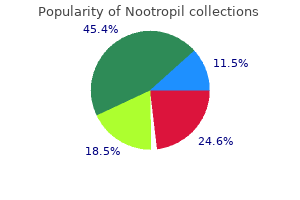
| Comparative prices of Nootropil |
| # | Retailer | Average price |
| 1 | Neiman Marcus | 535 |
| 2 | Army Air Force Exchange | 381 |
| 3 | SUPERVALU | 192 |
| 4 | A&P | 776 |
| 5 | Williams-Sonoma | 561 |
| 6 | OfficeMax | 901 |
| 7 | Brinker International | 845 |
Nootropil 800 mg buy discount line
Solubility of solids in liquids Solutions of solids in liquids are the commonest kind of resolution encountered in pharmaceutical practice symptoms 7 days before period nootropil 800 mg order amex. A pharmaceutical scientist should due to this fact pay consideration to the overall method of determining the solubility of a solid in a liquid and the various precautions that should be taken throughout such determinations medicine woman dr quinn 800 mg nootropil discount with mastercard. Determination of the solubility of a strong in a liquid the following points must be noticed in all solubility determinations: � the solvent and solute must be as pure as potential. The presence of small amounts of many impurities might both increase or lower the measured solubility. This is a particular downside with early preformulation samples, which are sometimes impure, and right here particular care should be taken. A saturated solution is obtained either by stirring excess powdered solute with solvent for several hours on the required temperature, until equilibrium has been attained, or by warming the solvent with an excess of the solute and permitting the combination to cool to the required temperature. It is crucial that some undissolved stable should be present at the completion of the cooling stage to ensure that the solution is saturated and never either subsaturated or supersaturated. A pattern of the saturated solution is obtained for evaluation by separating out undissolved strong from the answer. Filtration is often used, but precautions ought to be taken to ensure that: enhance the solubility and bioavailability of medicine, is given in Chapters 20 and 24. Temperature and heat input the dissolution process is often an endothermic one, i. In this type of system, provide of warmth will result in an increase within the solubility of a strong. Conversely, within the case of the much less generally occurring methods that exhibit exothermic dissolution, which try to evolve heat, a rise in supplied heat will end in a decrease in solubility. Plots of solubility versus temperature, referred to as solubility curves, are sometimes used to describe the effect of temperature on a given system. However, abrupt modifications in slope may be noticed with some systems if a change in the nature of the dissolving solid happens at a specific transition temperature. The solubility subsequently exhibits a change from a positive to a negative slope as the temperature exceeds the transition value, i. Membrane filters that can be utilized in conjunction with conventional syringes fitted with appropriate in-line adapters have proved to be successful. The quantity of solute contained in the pattern of saturated answer could additionally be decided by a selection of strategies. The selection of an appropriate method is affected by the nature of the solute and the solvent and by the focus of the answer. It also needs to be realized that even a small change within the molecular construction of a compound can have a marked effect on its solubility in a given liquid. For instance, the introduction of a hydrophilic hydroxyl group to a molecule can produce a large improve in water solubility. This is evidenced by the more than 100-fold greater aqueous solubility of phenol compared with benzene. In addition, the conversion of a weak acid to its sodium salt results in a much greater degree of ionic dissociation of the compound when it dissolves in water. The total interplay between solute and solvent is increased markedly and the solubility consequently rises. An instance of this effect is supplied by a comparability of the aqueous solubility of salicylic acid and that of its sodium salt, that are 1 in 550 and 1 in 1 respectively. The reduction in aqueous solubility of a parent drug by its esterification may be cited for example of the results of adjustments within the chemical structure of the solute. Such a discount in solubility may be useful to present an appropriate technique for: � masking the taste of a parent drug. This is achieved by using cosolvents such as ethanol or propylene glycol, that are miscible with water and which act as better solvents for the solute in query. The solubility of this drug can be elevated markedly by the incorporation of one or more water-miscible cosolvents so that an answer containing 500 mg in 10 mL (and thus suitable for parenteral administration within the treatment of anaerobic infections) may be obtained. Crystal characteristics: polymorphism and solvation When the circumstances under which crystallization is allowed to happen are diversified, some substances produce crystals during which the constituent molecules are aligned in several methods with respect to one another within the lattice construction. These different crystalline forms of the same substance, which are often identified as polymorphs, consequently possess different lattice energies, and this difference is mirrored by modifications in other properties. For instance, the polymorphic form with the bottom free power will be the most stable and possess the highest melting level. Other less stable (or metastable) varieties will tend to transform into essentially the most secure one at rates that depend on the power variations between the metastable and secure forms. Polymorphs are explained more totally in Chapter eight (which additionally contains a proof of why polymorphs may have completely different solubilities) and Chapter 23. Examples of the significance of polymorphism with respect to the bioavailability of medication are given in Chapter 20. The impact of polymorphism on solubility is particularly necessary from a pharmaceutical viewpoint, because it provides a method of accelerating the solubility of a crystalline material, and therefore its rate of dissolution, by using a metastable polymorph. Although the more soluble polymorphs are metastable and will convert to the secure kind, the speed of such conversion is commonly sluggish sufficient for the metastable form to be thought to be being sufficiently stable from a pharmaceutical viewpoint. There are merchandise on the market containing a extra soluble, but less stable, polymorph of the drug, the place the chosen polymorph is secure sufficient to survive the permitted storage conditions and declared shelf life. Examples of the significance of polymorphism with respect to the occurrence of crystal progress in suspensions are given in Chapter 26. In addition to the impact of polymorphism, the lattice constructions of crystalline supplies may be altered by the incorporation of molecules of the solvent from which crystallization occurred (discussed in Chapter 8). The resultant solids are referred to as solvates and the phenomenon is referred to appropriately as solvation. The alteration in crystal construction that accompanies solvation will affect the inner energetics of the solid such that the solubility of the solvated and unsolvated crystals will differ. Consequently, hydrated crystals tend to exhibit a decrease aqueous solubility than their unhydrated forms. Examples of the consequences of solvation and the attendant modifications in solubilities of medicine on their bioavailabilities are given in Chapter 20. Precipitation could happen, therefore, as a outcome of the solubility of the un-ionized species is normally lower than that of the ionized kind. Conversely, in the case of solutions of weakly primary drugs or their salts, precipitation is favoured by an increase in pH. Such precipitation is an instance of one kind of chemical incompatibility that could be encountered within the formulation of liquid medicines. This will have an effect on the degree of ionization of the drug molecules, which in flip influences their solubility and their capability to be absorbed. This side is discussed elsewhere in this e-book in some element, and the reader is referred specifically to Chapters three and 20.
Order nootropil 800 mg fast delivery
The depth of the secondary minimum is dependent upon the particle measurement medicine shoppe cheap 800 mg nootropil overnight delivery, and particles might must medications jamaica 800 mg nootropil generic visa be of radius 1 �m or greater earlier than the engaging drive is sufficiently great for flocculation to happen. An estimation of the floor potential may be obtained from zeta potential measurements. As could be seen, the repulsion energy is an exponential operate of the gap between the particles and has a variety of the order of the thickness of the double layer. For an meeting of molecules, dispersion forces are additive, summation leading to long-range attraction between colloidal particles. This latter means that there shall be an elevated tendency for particles to flocculate in the secondary minimal, and this is the precept of the controlled flocculation method to pharmaceutical suspension formulation described later. The main maximum may also be lowered (and the secondary minimum deepened) by including substances, such as ionic surface-active agents, which are particularly adsorbed inside the Stern layer. Here is decreased and hence the zeta potential; the double layer is often not compressed. The elements of a mix of hydrophilic colloids can subsequently be separated by a strategy of fractional precipitation, which involves the salting out of the varied parts at completely different concentrations of electrolyte. Lyophilic colloids could be thought of to become lyophobic by the addition of solvents corresponding to acetone and alcohol. The particles turn out to be desolvated and are then very sensitive to precipitation by added electrolyte. Coacerva- Stability of lyophilic methods Solutions of macromolecules, lyophilic colloidal sols, are stabilized by a combination of electrical double layer interaction and solvation, and each of those stabilizing components must be sufficiently weakened earlier than attraction predominates and the colloidal particles coagulate. Hydrophilic colloids are unaffected by the small amounts of added electrolyte which trigger hydrophobic sols to coagulate. However, when the focus of electrolyte is high, particularly with an electrolyte whose ions turn out to be strongly hydrated, the colloidal materials loses its water of solvation to these ions and coagulates, i. Variation within the diploma of solvation of different hydrophilic colloids affects the concentration of soluble electrolyte required to produce their tion is the separation of a colloid-rich layer from a lyophilic sol as the end result of the addition of one other substance. This layer, which is current within the type of an amorphous liquid, constitutes the coacervate. Simple coacervation could additionally be led to by a salting-out effect on addition of electrolyte or addition of a nonsolvent. Complex coacervation occurs when two oppositely charged lyophilic colloids are mixed. Gelatin at a pH beneath its isoelectric level is positively charged, and acacia above about pH 3 is negatively charged; a combination of solutions at about pH 4 ends in coacervation. If the coacervate is formed in a stirred suspension of an insoluble stable, the macromolecular material will encompass the stable particles. The coated particles may be separated and dried, and this system varieties the basis of one technique of microencapsulation. The coating protects the drug from chemical assault, and microcapsules could also be given orally to delay the action of the medicament. When added in small amounts, many polyelectrolyte and polymer molecules (lyophilic colloids) can adsorb concurrently onto two particles and are long sufficient to bridge the energy barrier between the particles. This can even occur with neutral polymers when the lyophobic particles have a high zeta potential (and would thus be considered a stable sol). Use is made from this property of small quantities of polyelectrolytes and polymers in removing colloidal material, resulting from sewage, in water purification. On the opposite hand, if larger amounts of polymer are added, adequate to cover the floor of the particles, then a lyophobic sol may be stabilized to coagulation by added electrolyte � the so-called steric stabilization or protecting colloid effect. Steric repulsion can be defined by reference to the free energy changes that happen when two polymer-covered particles work together. Here the impact of the entropy change opposes aggregation and outweighs the enthalpy term; that is termed entropic stabilization. Interpenetration Steric stabilization (protective colloid action) It has lengthy been known that nonionic polymeric materials such as gums, nonionic surface-active brokers and methylcellulose adsorbed at the particle surface can stabilize a lyophobic sol to coagulation even within the absence of a big zeta potential. Consequently, this effect is termed enthalpic stabilization and is common with aqueous dispersions, particularly where the stabilizing polymer has polyoxyethylene chains. The launched water molecules have larger degrees of freedom than these within the bound state. The particles hyperlink collectively to form an interlaced community, thus imparting rigidity to the construction; the continuous section is held throughout the meshes. Often solely a small proportion of disperse phase is required to impart rigidity; for example, 1% agar in water produces a agency gel. Clays corresponding to bentonite, aluminium magnesium silicate (Veegum) and to some extent kaolin kind gels by flocculation in a special method. They are hydrated aluminium (aluminium/magnesium) silicates whose crystal structure is such that they exist as flat plates. The forces holding the particles collectively in this type of gel are relatively weak � van der Waals seventy six forces within the secondary minimal flocculation of aluminium hydroxide, electrostatic attraction in the case of the clays. Because of this, these gels present the phenomenon of thixotropy, a nonchemical isothermal gel�sol�gel transformation. Flocculation in gels is the reason for his or her anomalous rheological properties (see Chapter 6). This phenomenon of thixotropy is used in the formulation of pharmaceutical suspensions. Gelation of lyophilic sols Gels formed by lyophilic sols could be divided into two teams depending on the character of the bonds between the chains of the network. Gels of type I are irreversible methods with a three-dimensional community formed by covalent bonds between the macromolecules. Typical examples of this sort of gel are the swollen networks that have been formed by the polymerization of monomers of water-soluble polymers in the presence of a cross-linking agent. Such polymers have been used in the fabrication of expanding implants that imbibe physique fluids and swell to a predetermined quantity. Implanted in the dehydrated state, these polymers swell to fill a physique cavity or give form to surrounding tissues. These gels are warmth reversible, a transition from the sol to gel occurring on either heating or cooling. Poly(vinyl alcohol) options, for example, gel on cooling to beneath a certain temperature referred to as the gel level. Because of their gelling properties, poly(vinyl alcohol)s are used as jellies for application of medication to the skin. On utility, the gel dries rapidly, leaving a plastic movie with the drug in intimate contact with the pores and skin. These compounds are amphiphilic and lots of kind micelles with a hydrophobic core comprising the polyoxypropylene blocks, surrounded by a shell of the hydrophilic polyoxyethylene chains.
800 mg nootropil order
In the previous medicine 20th century buy discount nootropil 800 mg, it has been thought of because the centre for auditory reflexes symptoms of hiv generic 800 mg nootropil visa, however the out there proof signifies that it helps in localizing the supply of sounds. White Matter Transverse Section of Midbrain on the Level of Superior Colliculi Grey Matter 1 the crus cerebri contains: a. Temporopontine, parietopontine and occipitopontine fibres within the lateral one-sixth. The lemnisci (medial, trigeminal, spinal and lateral) are arranged in the form of a band during which they lie within the order mentioned (from medial to lateral side) like a necklace. The medial longitudinal bundle lies in shut relation to the trochlear nucleus (somatic efferent column). It decussates in the superior medullary velum, and emerges lateral to the frenulum veli. Nucleus of oculomotor nerve with EdingerWestphal nucleus within the ventromedial half. It controls reflex movements of the eyes, and of the head and neck in response to visual stimuli. The pretectal nucleus is a crucial part of the pathway for light reflex and the consensual reflex. Its lesion causes Argyll Robertson pupil in which the light reflex is misplaced but lodging reflex stays intact. It receives afferents from the superior cerebellar peduncle, globus pallidus, subthalamic nucleus and cerebral cortex. It gives efferents to the spinal wire (rubrospinal tract), reticular formation, thalamus, olivary nucleus, subthalamic nucleus, and so forth. The identical lemnisci as seen within the decrease half apart from the lateral lemniscus which has terminated in the inferior colliculus. The decussation of the tectospinal and tectobulbar tracts varieties the dorsal tegmental decussation. The decussation of the rubrospinal tracts varieties the ventral tegmental decussation. These fibres reach as a lot as interstitial nucleus of Cajal on the higher end of aqueduct. Tremors and twitching of opposite side because of injury to purple nucleus and superior cerebellar peduncle. Features embody weak point of upward gaze and vertical nystagmus as a result of lesion of superior colliculus. The fibres of sunshine reflex take following course: Retina optic nerve optic chiasma optic tractsome fibres to pretectal nucleus of each side Edinger-Westphal nucleus3rd nerve nucleus and 3rd nerve ciliary ganglion short ciliary nerves pupil constricts. A person affected by syphilis complains of lack of ability to see in response to light thrown in the eyes, whereas he can learn and see close by things: � Where is the lesion Ans: In such circumstances, the light reflex is misplaced, whereas accommodation reflex is retained. Such a 1�10 From Medical Council of India, Competency based mostly Undergraduate Curriculum for the Indian Medical Graduate, 2018;1:44�80. Draw a labelled diagram of transverse section of medulla oblongata at the degree of sensory decussation. Temporopontine, parietopontine and occipitopontine fibres in lateral one-sixth half d. The anterior aspect of the cerebellum is marked by a large and deep notch during which the pons and medulla are lodged. It is restricted in front by the fissura prima (on the superior surface), and by the posterolateral fissure (on the inferior surface). Where both the superior and inferior surfaces of the cerebellum are drawn in a single aircraft. The higher part of the diagram, above the horizontal fissure, represents the superior floor; and the decrease part, beneath the horizontal fissure represents the inferior surface. It is made up of the anterior lobe (except lingula), and the pyramid and uvula of the inferior vermis. It is made up of the posterior/middle lobe (the largest a part of the cerebellum) besides the pyramid and uvula of the inferior vermis. It is primarily concerned with the regulation of nice actions of the physique (Table 6. Lateral Zone Connected with association areas of the brain and is concerned in planning, programming and coordination of muscular activities of the entire body. It is completed through dentato-rubro-thalamo-cortical tract, descending corticospinal tracts. Concerned with control of muscles of distal elements of limbs like hands and toes via rubrospinal tract. Planning and programming of purposeful and speedy actions including their period and termination � Acts as a suggestions centre between cerebral cortex and peripheral motor movements Spinocerebellum Dorsal spinocerebellar Ventral spinocerebellar Cuneocerebellar tract Reticulocerebellar Trigeminocerebellar Pontocerebellar tract Olivocerebellar Cerebelloreticular Cerebello-olivary Neocerebellum Dentatothalamic Dentatorubral Table 6. Inferior cerebellar peduncle (connects cerebellum to medulla oblongata) Afferent tracts 1. Posterior spinocerebellar Cuneocerebellar (posterior external arcuate fibres) Olivocerebellar Parolivocerebellar Reticulocerebellar Vestibulocerebellar Anterior exterior arcuate fibres Striae medullaris Trigeminocerebellar 1. This is in marked contrast to cerebrum which controls the alternative half of the body. It controls and coordinates these by affecting agonists, antagonists and synergists. Spinocerebellum, vermis and intermediate areas receive afferents from motor cortex via corticopontocerebellar fibres. All sensory information of muscles, joints, cutaneous, auditory and visible elements are relayed here. It corrects and modifies ongoing movements through thalamocortical projections, reticulospinal and rubrospinal tracts. These patients show inattention, grammatical errors in speech and patchy reminiscence loss. Intention tremors (tremors only throughout movements) tested by finger-nose and heel-knee exams. Adiadochokinesia which is incapability to perform rapid and regular alternating movements, like pronation and supination. Nystagmus is to and fro oscillatory actions of the eyeballs while looking to either facet. The cortex incorporates three layers: 1 Molecular layer: It consists of unmyelinated nerve fibres which are derived from the parallel fibres of axons of granule cells, axons of stellate and basket cells, sensory climbing fibres, dendrites of Purkinje and Golgi cells. Each fibre gives collateral branches to synapse with deep cerebellar nuclei and make monosynaptic contacts after coiling around the non-spinous a part of the dendritic tree of 1 Purkinje cell. These neurons migrate dorsally and type the rhombic lip which forms the cerebellum. In its centre, the paleocerebellum develops, splitting the archicerebellar parts into two parts-the lingula and flocculonodular lobe.
Syndromes
- Stone in salivary duct
- Washing of the skin (irrigation) -- perhaps every few hours for several days
- Assistive devices, such as special eating utensils, wheelchairs, bed lifts, shower chairs, walkers, and wall bars
- Disease that results in breakdown of muscle fibers (rhabdomyolysis)
- Insist that others wash their hands with warm water and soap before touching your baby.
- Fever
- 7 - 12 months: 400 IU (10 mcg/day)
- Chronic back pain
- Swollen or tender lymph nodes (lymphadenopathy)
Buy nootropil 800 mg on-line
Venous Drainage In the broad ligament symptoms kidney failure dogs nootropil 800 mg with mastercard, uterine artery is anterosuperior to ureter by 2 symptoms 9dp5dt discount 800 mg nootropil free shipping. Ureter runs forwards barely above the lateral fornix of vagina and is 2 cm lateral to supravaginal part of cervix. Left ureter to usually extra close to vagina, as uterus is slightly on the right facet and vagina is on the left side of median airplane. One has to watch out of ureter, particularly during ligation of the uterine artery. Nerve Supply Ureter in Female Pelvis As ureter lies anterior to internal iliac artery and immediately behind ovary, it forms the posterior boundary of the ovarian fossa. In its anteromedial part, ureter is related to the uterine artery, cervix and vaginal fornices. Ureter lies in endopelvic fascia in inferomedial part of broad ligament of uterus, where it could be damaged. Sympathetic nerves are vasoconstrictor and parasympathetic nerves are vasodilator. It is kept moist by the cervical glands from above and the higher vestibular glands from beneath. Inspection: Vagina is first inspected at the introitus by separating the labia minora with the left hand. Next the speculum examination is done to examine the cervix and vaginal vault, and to take the vaginal swab. With the examining fingers, one can really feel: � Anteriorly, the urethra, bladder and pubic symphysis; � Posteriorly, the rectum and pouch of Douglas; � Laterally, the ovary, tube, lateral pelvic wall, thickened ligaments, and ureters; and � Superiorly, the cervix. Bimanual (abdominovaginal) examination helps in the assessment of the dimensions and place of the uterus, enlargements of the ovaries and tubes, and the opposite pelvic masses. Vaginitis is often caused by the trichomonas, monilial and gonococcal infections. Similarly, trauma to the anterior and posterior partitions of vagina could cause the vesicovaginal, urethrovaginal and rectovaginal fistulae. Various elements of ovary are germ cells, follicular cells and the stromal cells. Follicular cells are derived from the epithelial cells of the coelomic epithelium, while stromal cells are derived from mesoderm. Its descent is interrupted because of the presence of the one uterus, which divides the gubernaculum into ligament of ovary and round ligament of uterus. Epoophoron It consists of 10 to 15 parallel tubules located within the lateral part of the mesosalpinx between the ovary and the uterine tube. Paroophoron It consists of some very short rudimentary tubules situated within the broad ligament between the ovary and Vesicular Appendix Occasionally, one or two pedunculated cysts are discovered hooked up to the fimbriated end of the uterine tube. These Section Occasionally, the duct of the epoophoron is way bigger than ordinary and is called the duct of Gartner. It may be traced first along the uterine tube, after which along the lateral margin of the uterus up to the extent of the interior os. Further down, it runs via the cervix and the lateral wall of the vagina, and ends close to the free margin of the hymen. M�llerian duct proliferates due to presence of oestrogens and absence of testosterone and of m�llerian inhibiting substance. The distal parts of the two ducts fuse to type the one uterovaginal canal which supplies rise to uterus with its fundus, body and cervix components. Wolffian duct or mesonephric duct forms the trigone of urinary bladder as useful component. The medulla incorporates hilus cells, homologous to the interstitial cells of the testis. Endoderm on either side of M�llerian tubercle proliferates to type two sinovaginal bulbs which fuse to form vaginal plate. It opens through an endodermal partial septum-the hymen within the definitive urogenital sinus. External Genitalia Mesenchymal cells migrate round cloacal membrane to kind cloacal folds. Cloacal folds get divided into urethral folds anteriorly and anal folds posteriorly. This occurs at the same time that the cloacal membrane gets divided into urogenital membrane and anal membrane. Lateral to urethral folds, another pair of folds, the genital swellings make their look. Genital tubercle varieties clitoris; urethral folds form labia minora, genital swellings type labia majora, urogenital membrane gets ruptured to form the vestibule. Mnemonics Due to repeated childbirths, a female felt one thing out at the perineum, especially while standing. Its helps are: � Muscular: All parts of levator ani, perineal physique with its hooked up ten muscle tissue and distal urethral sphincter mechanism. A chapter that provides a detailed evaluate of the helps of the vagina and the pelvic organs. A landmark study that prospectively recognized levator trauma after vaginal supply utilizing non-invasive three-dimensional ultrasound. Neuroanatomy of the female abdominopelvic area: A review with utility to pelvic ache syndromes. A detailed review of the somatic and autonomic nerve supply of the feminine genital system and feminine pelvis. The exterior genitalia are the penis and the scrotum containing testis, epididymis and part of ductus deferens. The internal genitalia on both sides are, the a part of ductus deferens, the seminal vesicle, the ejaculatory ducts, and single prostate gland. The exterior genitalia have been described in Chapter 17, and the male urethra in Chapter 30. Follow it from there because it hooks round the lateral side of inferior epigastric artery to cross backwards and medially across the external iliac vessels to enter into the lesser pelvis. Follow the deep dorsal vein of the penis and its two divisions into the prostatic venous plexus situated within the angle between the bladder and the prostate. Location and Course In its course, the vas lies successively: 1 Within the scrotum along the posterior border of the testis. It runs upwards to the superficial inguinal ring, and then traverses the inguinal canal. In the vertical part of its course, it can be felt as a cord-like structure inside the spermatic wire. Here it crosses the obliterated umbilical artery, the obturator nerve and vessels and the vesical vessels. It then crosses the ureter and bends medially at proper angles, to enter the sacrogenital fold of peritoneum.
Order nootropil 800 mg overnight delivery
Particle size instrumentation is growing quickly medicine 7767 cheap nootropil 800 mg amex, however a abstract of the rules of the strategies most commonly encountered in pharmaceutics is introduced right here medicine - nootropil 800 mg discount visa, primarily based on the necessary thing options of each approach. Principles of measurement Sieve evaluation uses a woven, punched or electroformed mesh, typically produced from stainless-steel or brass, with identified aperture dimensions which forms a bodily barrier to the particles. A sieve stack often contains six to eight sieves with an aperture progression based mostly on a 2 change in space between adjacent sieves. Powder is loaded onto the coarsest sieve on the top of the assembled stack, and the nest is subjected to mechanical agitation. After an appropriate time, the sieve diameter of a particle is the length of the side of the minimum sq. aperture through which it has handed. The weight of material collected on every stage is determined and used to plot a cumulative-undersize plot. In follow, sieves can be obtained for dimension evaluation over a range from 5 �m to one hundred twenty five 000 �m. Sample preparation and analysis situations Sieve evaluation is usually carried out with powders in the dry state, though for powders in liquid suspension or for these which agglomerate during dry sieving, a process of moist sieving can be utilized. Sample preparation and evaluation situations Specimens ready for mild microscopy have to be adequately dispersed on a microscope slide to keep away from evaluation of agglomerated particles. Specimens for scanning electron microscopy are ready by their being mounted to aluminium stubs earlier than being sputter coated with a movie of gold a number of nanometres in thickness. Alternative strategies Another type of sieve evaluation, known as air-jet sieving, makes use of particular person sieves quite than a whole nest of sieves. The course of starts with the finest-aperture sieve and progressively removes the undersize particle fraction by sequentially increasing the apertures of each sieve, encouraging particles to cross through each aperture underneath the influence of a partial vacuum applied beneath the sieve mesh. A reverse air jet circulates beneath the sieve mesh, blowing oversize particles away from the mesh to prevent blockages. Air-jet sieving is usually extra environment friendly and reproducible than typical mechanically vibrated sieve analysis, though with finer particles, agglomeration can turn out to be a problem. In the associated technique, sonic-sifter sieving, comparatively small powder samples are lifted in a vertically oscillating column of air, such that particles are carried in opposition to a sieve mesh at a set variety of pulses per minute. Light microscopy Principles of measurement Size evaluation by gentle microscopy is carried out on two-dimensional images of particles which are generally assumed to be randomly oriented in three dimensions. In many cases, this assumption is valid, although for crystal dendrites, fibres or flakes, it is very inconceivable that the particles will orient with their minimum dimensions in the airplane of measurement. Under such situations, measurement analysis is performed accepting that such particles are considered in their most stable orientation. This will lead to an overestimation of dimension as a end result of the bigger dimensions of the particle shall be noticed, because the smallest dimension will most often orient vertically. The two-dimensional photographs are analysed in accordance with the specified equivalent diameter. With use of a conventional mild microscope, particle dimension evaluation could be carried out with an eyepiece graticule which has previously been calibrated. One also can use a graticule which has a collection of opaque and transparent circles of different diameters, usually in a 2 progression. The field of view is split into segments to facilitate measurement of different numbers of particles. Both scanning electron microscopy evaluation and transmission electron microscopy evaluation permit the decrease particle sizing restrict to be significantly extended over that attainable with a light-weight microscope. Sample preparation and analysis conditions Particle measurement distributions can be determined by examination of a powder as it sediments in a liquid. If the powder is hydrophobic, it could be essential to add a dispersing agent to assist wetting. In circumstances the place the powder is soluble in water, it will be necessary to use nonaqueous liquids or carry out the analysis in a gasoline. Image evaluation With manual microscopy, only a few particles can be examined and sized in an inexpensive time. This dangers selection of an unrepresentative sample, operator subjectivity and operator fatigue. Automated picture analysis, for gentle and electron microscopy, has the advantages of being more objective and far sooner than manual evaluation, and it additionally permits a a lot wider number of measurement and shape parameters to be processed. Image evaluation may be static, whereby particles on a microscope slide are inspected with use of a microscope and digital digicam, or dynamic (flowimage analysis), whereby pictures of particles dispersed in a liquid are captured by a camera as they cross through a move cell. Principles of measurement the strategies of dimension analysis by sedimentation could be divided into two primary categories according to the tactic of measurement used. One type is based on the measurement of particles in a retention zone; a second type makes use of a nonretention measurement zone. An instance of a nonretention zone measurement method is named the pipette methodology. In this technique, known volumes of suspension are withdrawn and the concentration variations are measured with respect to time. The Andreasen fixed-position pipette contains a graduated cylinder which can maintain roughly 500 mL of suspension fluid. A pipette is located centrally within the cylinder and is held in place by a ground-glass stopper so that its tip coincides with the zero degree. A three-way tap permits fluid to be drawn into a ten mL reservoir, which may then be emptied right into a beaker or centrifuge tube. The amount of powder may be decided by weight following drying or centrifuging; alternatively, chemical analysis of the collected particles could be performed. Sedimentation methods Equivalent sphere diameters (Frictional) drag diameter, dd, and Stokes diameter, dSt (Table 9. Drag is certainly one of three forces performing on a particle sedimenting in a gravitational field. A drag drive, Fd, acts upwards, as does a buoyancy drive, Fb; a third drive is gravity, Fg, which acts because the driving force of sedimentation. At the constant terminal velocity, which is quickly achieved by sedimenting particles, the drag pressure becomes synonymous with particle movement. Thus for a sphere of diameter d and density s, falling in a fluid of density f, the equation of movement is Fd = (s - f)Fg d three 6 (9. In this methodology the quantity of sedimented particles falling onto a stability pan suspended in the fluid is recorded. The continual improve within the weight of sediment is recorded with respect to time. Alternative strategies One of the limitations of gravitational sedimentation is that beneath a diameter of approximately 5 �m, particle settling turns into extended and is subject to interference from convection, diffusion and Brownian motion. One can decrease these effects by rising the driving drive of sedimentation by replacing gravitational forces with a bigger centrifugal drive. To decrease the impact of distance on the sedimenting drive, a twolayer fluid system can be used. A small quantity of concentrated suspension is launched onto the floor of a bulk sedimentation liquid often known as the spin fluid. With use of the technique of disc centrifugation, all particles of the same dimension are in the identical place in the centrifugal field and hence move with the identical velocity. An adaptation of a retention zone gravity sedimentation technique is called a micromerograph and measures sedimentation of particles in a gas rather than a fluid.
Buy nootropil 800 mg otc
Patella (knee cap) is the biggest sesamoid bone of the body medications with gluten nootropil 800 mg cheap on-line, developed within the tendon of quadriceps femoris treatment for hemorrhoids 800 mg nootropil discount overnight delivery. A small nick is given in the femoral artery and cannula introduced so that its tip factors towards the top end and 8. The areas throughout the interrupted lines have a well-defined membranous layer of superficial fascia Lower Limb It is easily seen and felt in entrance of the knee. Tibial tuberosity is a blunt prominence in entrance of the higher end of tibia, marking the upper finish of the shin. The medial and lateral condyles of the femur and of the tibia form giant bony plenty on the sides of the knee. The most outstanding points on the perimeters of the femoral condyles are referred to as the medial and lateral epicondyles. Vastus medialis varieties a fleshy prominence above the medial condyle of femur, notably in an extended knee. Adductor tubercle is a bony projection from the uppermost a half of the medial condyle of femur to which the tendon of adductor magnus is connected. To palpate the tubercle, flex the knee partly and note the extensive, shallow groove that seems posterior to the mass of vastus medialis. The superficial fascia has two layers, a superficial fatty layer and a deep membranous layer, which are steady with the corresponding layers of the anterior abdominal wall. The two layers are most distinct in the uppermost part of the thigh, near the groin, the place the cutaneous nerves, vessels and lymph nodes lie between the 2 layers. When the urethra is injured within the perineum, urine might flow out or extravasate into the interval deep to the membranous layer of superficial fascia. This urine can cross up into the anterior stomach wall from the place it may possibly enter the upper a half of the thigh. The superficial fascia incorporates cutaneous nerves, cutaneous arteries, the good saphenous vein and its tributaries, and the superficial inguinal lymph nodes. The presence of few stitches signifies that embalming for preservation of the physique has been carried out from this web site. Procedure for embalming: A 6 cm lengthy vertical incision is given in the higher medial aspect of thigh. After reflecting pores and skin and fasciae, femoral sheath is incised to visualise the femoral artery. The intermediate cutaneous nerve of thigh (L2, 3) is a branch of the anterior division of the femoral nerve. It pierces the deep fascia on the junction of the higher onethird and middle one-third of the thigh. It divides into two or extra branches and provides a strip of skin on the entrance of the thigh extending from the sartorius to the knee. The medial cutaneous nerve of the thigh (L2, 3) is a department of the anterior division of the femoral nerve. The nerve supplies the pores and skin on the medial side of the decrease twothirds of the thigh. The saphenous nerve (L3, 4) is a department of the posterior division of the femoral nerve. Before piercing the deep fascia the saphenous nerve offers off the infrapatellar department which runs downwards and laterally, and provides the pores and skin over the ligamentum patellae. Patellar Plexus Patellar plexus is a plexus of fantastic nerves located in front of the patella, the ligamentum patellae and the higher end of the tibia. It is fashioned by contributions from: 1 the anterior division of the lateral cutaneous nerve 2 the intermediate cutaneous nerve three the anterior division of the medial cutaneous nerve four the infrapatellar branch of the saphenous nerve. Cutaneous Arteries the lateral cutaneous nerve of thigh (L2, 3) is a branch of the lumbar plexus. It emerges behind the lateral end of the inguinal ligament, divides into anterior and posterior branches, and provides the pores and skin on the this is the largest and longest superficial vein of the lower limb (Saphes = easily seen). In the thigh, it inclines forwards to attain the saphenous opening where it pierces the cribriform fascia and opens into the femoral vein. Superficial Inguinal Lymph Nodes the superficial inguinal lymph nodes are variable in their number and measurement. Their association is T-shaped, there being a decrease vertical group and an upper horizontal group. Give a curved incision across the scrotum/pudendal cleft in direction of upper medial side of thigh. Extend it vertically down beneath the medial condyle of tibia until the extent of tibial tuberosity. Reflect the skin laterally, exposing the superficial fatty and deeper membranous layers of superficial fascia. Identify the nice saphenous vein within the medial a half of anterior floor of thigh. Draining into its higher part are its three superficial tributaries, specifically superficial circumflex iliac, superficial epigastric and superficial exterior pudendal. The vertical group of superficial inguinal lymph nodes lie along the higher a half of nice saphenous vein. Dissect the superficial inguinal ring 1 cm above and lateral to the pubic tubercle. Trace the good saphenous vein backwards till it pierces the specialised deep fascia often identified as cribriform fascia to drain into the femoral vein enclosed within the femoral sheath. Bursae are lubricating mechanisms which are supplied at sites of friction to smoothen motion. Prepatellar Bursa Prepatellar bursa lies in entrance of the lower a half of the patella and of the upper a half of the ligamentum patellae. The fascia lata is a troublesome fibrous sheath that envelops the whole of the thigh like a sleeve. Inferiorly, on the front and sides of the knee, the fascia lata is attached to subcutaneous bony prominences and the capsule of the knee joint. Posteriorly, it forms the sturdy popliteal fascia which is steady below with the fascia of the again of the leg. The superficial, lamina is connected to tubercle of iliac crest, and deep lamina to the capsule of hip joint. Inferiorly, the tract is hooked up to a clean area on anterior surface of the lateral condyle of tibia. Two important muscles are inserted into its higher half, between the superficial and deep laminae. These are the three-fourths part of the gluteus maximus; and the tensor fasciae latae.
Order 800 mg nootropil mastercard
The roof medicine klimt 800 mg nootropil with visa, shaped by the orbital plate of the frontal bone and medications errors discount nootropil 800 mg with mastercard, posteriorly, the lesser wing of the sphenoid, separates the orbit from the anterior cranial fossa. The lateral wall, formed by the zygomatic bone and the higher wing of the sphenoid, separates the orbit from the temporal fossa. The medial wall consists of, from anterior to posterior, the maxilla, the lacrimal bone, the orbital plate of the ethmoid and the body of the sphenoid. The orbital cavity has three posterior openings, the superior and inferior orbital fissures laterally and the optic canal medially. The lateral part transmits the lacrimal, trochlear and frontal nerves, the medial, superior and inferior divisions of the oculomotor nerve, the nasociliary nerve and the abducent nerve. The inferior orbital fissure, between the floor and lateral wall, opens into the pterygopalatine fossa medially and the infratemporal fossa laterally. It transmits the maxillary nerve, which in the canal turns into the infraorbital nerve, and its zygomatic branch, together with speaking veins. The optic canal opens into the apex of the cavity and conveys the optic nerve and ophthalmic artery from the middle cranial fossa. A fracture of the medial wall might involve the ethmoidal and sphenoidal sinuses; a fracture of the decrease margin may contain the maxillary sinus and infraorbital nerve, leading to lack of sensation over the cheek. The pigmented choroid, which, anteriorly, has a circular thickening, the ciliary physique, containing the ciliary muscle. In front of the ciliary body the choroid thins to kind the iris, with its central aperture, the pupil. The iris incorporates the circular sphincter pupillae and the radial dilator pupillae muscular tissues. The retina accommodates the sunshine receptors, which relay impulses along the optic nerve. About three mm lateral to the disc, on the visual axis, is a small depression, the fovea centralis. The biconvex lens divides the eyeball cavity right into a posterior part filled with the jelly-like vitreous physique and an anterior part crammed with the extra fluid aqueous humour. The iris additional divides the anterior half into an anterior chamber between the iris and cornea and a posterior chamber between the iris and lens. Accommodation, the process by which close to objects are focused on the retina, is achieved by contraction of the ciliary muscle drawing the ciliary physique forwards and stress-free the suspensory ligament. Occlusion of the central artery of the retina could happen as the outcomes of arterial illness or because of embolism (the trapping of free-floating clots) in the artery. Ciliary nerves carry autonomic fibres to the muscles of the iris and the ciliary muscle tissue and sensory fibres from the conjunctiva; the long ciliary nerves carry sympathetic fibres and arise from the nasociliary nerve. The quick ciliary nerves come up from the ciliary ganglion and carry postganglionic parasympathetic fibres. The sensory part is the optic nerve, and the motor component includes parasympathetic fibres carried throughout the oculomotor nerve. Thickenings of the fascia connected to the lacrimal and zygomatic bones type the medial and lateral verify ligaments of the eye, and a thickening under the eyeball varieties the suspensory ligament that provides a hammock-like assist for the eye. Anteriorly the muscles move forwards, in positions implied by their names, to be connected to the sclera just in entrance of the equator of the eyeball. It then passes backwards and laterally to acquire attachment to the posterolateral surface of the eyeball, behind the equator. The inferior oblique is hooked up anteriorly to the anteromedial floor of the orbit and passes backwards to be attached to the posterolateral floor of the eyeball, behind the equator. Nerve provide Lateral rectus is served by the abducent nerve, superior indirect by the trochlear nerve and the remaining muscles by the oculomotor nerve. Looking left includes contraction of the left lateral rectus and right medial rectus; trying down and to the proper includes the left superior indirect and the proper inferior rectus; looking up and to the left involves the left inferior oblique and the proper superior rectus. Oculomotor palsy, when complete, results in paralysis of a lot of the eye muscular tissues, along with levator palpebrae superioris and the sphincter pupillae. The palsy could end result from viral an infection, however occasionally it may be a non-localizing sign of increased intracranial pressure (because the long intracranial course of the nerve renders it particularly susceptible to stretching by the raised pressure). Trochlear nerve palsy is rare; sufferers attempt to reduce the diplopia it causes by tilting the pinnacle. It enters the orbit via the optic canal, and gives ciliary branches, the central artery of the retina and muscular branches earlier than ending behind the medial facet of the higher eyelid by dividing into supratrochlear, supraorbital and dorsal nasal branches. Superior and inferior ophthalmic veins drain the orbit and pass by way of the superior orbital fissure to the cavernous sinus. They talk by way of the inferior orbital fissure with the pterygoid venous plexus posteriorly and with the facial vein anteriorly near the medial angle of the eye. Within the medial angle of each eyelid is a pink elevation, the lacrimal caruncle. Each eyelid has 5 layers: Levator palpebrae superioris Levator palpebrae superioris raises the higher eyelid. It is hooked up posteriorly above the frequent tendinous origin and anteriorly to the conjunctiva and tarsal plate. When the lids are closed the conjunctiva encloses a slim sac containing a small quantity of lacrimal fluid. The higher and decrease pouches between the lids and the eyeball are generally known as the conjunctival fornices. The eyelashes emerge via the pores and skin of the lid margin, and small sebaceous glands are associated with them. Eye closing is effected by orbicularis oculi, and the higher lid is raised by levator palpebrae superioris. The rash of herpes zoster (shingles) infection of the Vth cranial nerve will produce corneal ulcers. They comprise the lacrimal gland and its ducts, the conjunctival sac, the lacrimal sac and the nasolacrimal duct. The front of the eyeball and its conjunctival covering is continually washed by tears which would possibly be important to ensure the conjunctiva remains moist and viable. The lacrimal gland is a serous gland within the superolateral angle of the orbit behind the upper lid. It is almond-shaped, with a palpebral course of between the conjunctiva and the tarsal plate, and it has 6�12 ducts opening into the superior conjunctival fornix. It is equipped by a department of the ophthalmic artery and innervated by the facial nerve by way of the pterygopalatine ganglion; postganglionic parasympathetic secretomotor fibres cross in the lacrimal and zygomatic nerves. Each lacrimal canaliculus is about 10 mm lengthy and passes from the lacrimal punctum in every eyelid to the lacrimal sac. This is a skinny fibrous sac on the medial facet of the orbit within the lacrimal fossa; it receives both lacrimal canaliculi and drains to the nasolacrimal duct. The duct descends within the medial wall of the orbit and opens into the inferior meatus of the nasal cavity.
800 mg nootropil order with mastercard
All these arteries attain the entrance of the knee and participate in forming the anastomoses around the knee medicine over the counter nootropil 800 mg buy with amex. In coarctation of the aorta fungal nail treatment purchase nootropil 800 mg on line, the popliteal stress is lower than the brachial stress. Sudden occlusion of the artery could cause gangrene as much as the knee, however this is usually prevented by the collateral circulation through the profunda femoris artery. This may be a source of continuous traction or stretching on the artery, inflicting primary thrombosis of the artery in young topics. Branches 1 Three genicular or articular branches come up within the upper a half of the fossa. Superior medial genicular nerve lies above the medial condyle of femur, deep to the muscle tissue. Inferior medial genicular nerve lies along the higher border of popliteus and reaches inferior to the medial condyle of tibia. It supplies the pores and skin of decrease half of back of leg and complete of lateral border of the foot until the tip of little toe. The nerve to the popliteus crosses the popliteal artery, runs downwards and laterally, winds round the decrease border of the popliteus, and supplies it from its deep (anterior) floor. In addition to the popliteus, the nerve additionally provides the tibialis posterior, the superior tibiofibular joint, the tibia, the interosseous membrane, and the inferior tibiofibular joint. It extends from the superior angle of the fossa to the lateral angle, along the medial border of the biceps femoris. Superior lateral genicular nerve accompanies the artery of the same name and lies above the lateral femoral condyle. Recurrent genicular nerve arises the place common peroneal nerve divides into superficial and deep peroneal nerves. Sensory loss: Loss of sensation on complete of sole of foot, plantar side of digits and nail beds on dorsum of foot. It lies in the identical superficial airplane as the tibial these lie deep to the deep fascia close to the termination of the small saphenous vein. They obtain afferents from lateral part of sole, both superficial and deep components of back of leg and knee joint. It may get entrapped between the attachments of peroneus longus to the top and shaft of fibula. The superficial part lies partly in the superficial fascia across the patella and partly in fat behind the ligamentum patellae. The deep half lies on the femur and the tibia throughout their adjoining articular surfaces. Circumflex fibular from posterior tibial the medial and lateral arteries type longitudinal anastomoses on all sides of the patella. The longitudinal anastomoses are interconnected to type transverse anastomoses in relation to the patella and above the tibial tuberosity. It gives genicular branches in the higher half, cutaneous department in the middle half and muscular branches within the decrease part of the fossa. Ans: the blood strain in decrease limb is taken by auscultating the deeply placed popliteal artery. Surgical anatomy of the sural and superficial fibular nerves with an emphasis on the method to the lateral malleoulus. Relation of popliteal vessels to tibial nerve in numerous parts of popliteal fossa. It is incompletely separated from the medial compartment by the poorly defined posterior intermuscular septum. Identify its branches in back of thigh to every of the hamstring muscular tissues together with often for the brief head of biceps femoris muscle. Separate the hamstring muscle tissue to expose the ischial a half of the composite or hybrid adductor magnus muscle. The adductor magnus reaches solely as much as the adductor tubercle of the femur, however is included amongst the hamstrings because the tibial collateral ligament of the knee joint morphologically is the degenerated tendon of this muscle. The ligament is connected to medial epicondyle, two millimeters from the adductor tubercle. Lower part of the inferior ramus of the pubis Into the groove on the posterior surface of the medial condyle of the tibia. Weak extensor of the hip Chief flexor of the knee and medial rotator of the leg in semiflexed knee. Weak extensor of the hip Chief flexor of the knee and lateral rotator of leg in semiflexed knee. Weak extensor of the hip Adductor half causes adduction of thigh; Ischial part helps in extension of hip and flexion of knee 2. Course and Relations 1 In the pelvis: the nerve lies in front of the piriformis, beneath cover of its fascia. It runs downwards with a slight lateral convexity, passing between the ischial tuberosity and the larger trochanter. It runs vertically downwards up to the superior angle of the popliteal fossa, on the junction of the upper two-thirds and decrease one-third of the thigh, where it terminates by dividing into the tibial and the frequent peroneal nerves. It begins within the pelvis and terminates at the superior angle of the popliteal fossa by dividing into the tibial and common peroneal nerves. The sciatic nerve is accompanied by a small companion artery-arteria nervi ischiadici. Branches Lower Limb 1 Articular branches to the hip joint arise in the gluteal area. When a person sits on the sting of a tough table/chair, the nerve gets compressed between the edge of desk and femur. Pain normally begins in the gluteal region, and radiates alongside the back of the thigh, and the lateral side of the leg, to the dorsum of the foot. This is usually because of compression of a quantity of nerve roots forming the sciatic nerve. The lateral circumflex femoral branch of the profunda femoris divides into ascending, transverse and descending branches. The main provide to the again of the thigh is thru the perforating branches of the profunda femoris. Perforating Branches of the Profunda Femoris Artery Section the profunda femoris artery offers off four perforating arteries. These longitudinal anastomotic chains are fashioned by the branches of inner iliac, the femoral and popliteal arteries. This is formed by the companion artery of the sciatic nerve and the perforating arteries.
