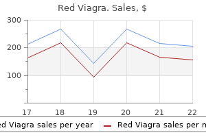
Red viagra 200 mg buy generic on line
If no specific leak is documented erectile dysfunction caused by ssri purchase red viagra 200 mg online, and collaterals are noted to drain into the venous system erectile dysfunction tips generic 200 mg red viagra amex, medical administration has a a lot higher success fee. If the duct may be identified, then transabdominal coil embolization has been successful. A persistent area (especially after pneumonectomy), widespread disruption (after esophagectomy for example), or persistent high output with medical therapy is associated with a particularly excessive failure rate, and earlier intervention is warranted. The website may be instantly visualized, in which case direct ligation (usually with pledget sutures) or glue application ought to be used. Localization could be assisted by feeding the affected person cream just prior to operation. Mass ligation at the degree of the diaphragm on the right aspect can resolve both right and left leaks. It is critical to acknowledge that the duct and surrounding tissue could be very friable, and thus ligation can result in one other website of leak. We have thus discovered that a critical component is to assure full decortication (to enable lung expansion), pleural abrasion or decortication, and if in doubt, proceed air flow for 24 hours to assist full lung expansion. Fibrothorax As talked about within the dialogue of retained hemothorax, symptomatic fibrothorax is more feared than real. It can occur following penetrating accidents to the thoracic inlet, after transmediastinal accidents, or after blunt trauma. Chylothorax can manifest in a delayed style with recurrent effusions, as persistent milky pleural output, or rarely as a pressure chylothorax. Chylothorax is more commonly seen as a complication following repair of aortic injury or esophageal resection. The diagnosis can be established by documenting triglyceride levels higher than 110 mg/dL and predominant lymphocytes within the effusion. If noted acutely, you will want to consider the potential for associated harm to adjacent structure, especially esophagus or aorta. Although low-fat diets do reduce the circulate of chyle, even oral water has been noted to improve chyle move. If the patient is, however, requiring vasopressors due to septic shock, parenchymal resection should be delayed, using percutaneous drainage or thoracoscopy. This ought to prompt consideration for thoracoscopic drainage early earlier than vigorous adhesions develop. In the absence of lung necrosis, either airway stents or direct reconstruction leads to good outcomes. Large, central, and irregular fragments must be removed 2 to three weeks after damage to permit surrounding inflammation to resolve. Asensio, Patrizio Petrone, Alejandro Perez-Alonso, Zachary Torgersen, Brian Biggerstaff, Brittney J. Ambrose Par�, the well-known French trauma surgeon, described two cases of penetrating cardiac accidents, both detailed from post-mortem studies. Wolf, in 1642, was the first to describe a healed wound of the guts, and Senac, in 1749, concluded that though all wounds of the guts have been serious, some wounds might heal and never be fatal. Larrey was the primary to describe the surgical strategy to the pericardium to relieve a pericardial effusion and is credited with pioneering the method for pericardial window. Billroth, in 1875 and in 1883, proclaimed his robust resistance to any attempt at cardiac damage repair. Block, in 1882, created cardiac wounds in a rabbit model and was profitable in attaining repair, thus demonstrating profitable recovery and suggesting that the same techniques could be applicable to humans. Also, Del Vecchio demonstrated cardiac damage healing after suturing the guts in a canine model. However, it took the braveness of Cappelen from Norway to try cardiac damage restore in a human; in 1895 he repaired a 2-cm left ventricular laceration together with ligation of a big branch of the distal left anterior descending coronary artery. This was adopted by Farina in Italy in 1896, who also attempted to repair a left ventricular wound; however, each sufferers succumbed. Rehn in Germany in 1896 was profitable in repairing a wound of the proper ventricle, and Hill, in 1902, was the first surgeon in the United States to efficiently repair a left ventricular harm. Duval described the median sternotomy incision, and Spangaro, in 1906, described the left anterolateral thoracotomy incision. Peck in 1909 was the first to describe profitable restore of a stab wound of the best atrium, and he reported a total of eleven patients. Smith was the first to develop a complete plan for cardiac injury administration, and for the primary time pointed out the risks of dysrhythmias occurring throughout cardiac manipulation. He additionally described the utilization of an Allis clamp near the apex to stabilize and hold the heart during suture placement. Beck in 1942 described the technique of putting mattress sutures underneath the bed of the coronary arteries. During the same 12 months, Griswold refined the strategies in the management of cardiac accidents and beneficial that every giant basic hospital should have available a sterile set of instruments plus an available working room 24 hours a day. Elkin in 1944 really helpful the administration of intravenous infusions before operation and pointed to the useful effects of increasing blood volume and thus cardiac output. These hallmark contributions have made it potential for sufferers sustaining penetrating cardiac injuries to survive today. Incidence Feliciano et al in 1983 described a 1-year expertise consisting of 48 cardiac injuries at Ben Taub Hospital in Houston. Mattox and associates in 1989 described a 30-year experience from the identical establishment reporting 539 cardiac accidents (18 cardiac accidents per year). Thus, penetrating cardiac accidents are uncommon and are usually seen solely in busy urban trauma facilities. According to a latest evaluate, 63% of all reported cardiac accidents in America are brought on by gunshot wounds and 36% are attributable to stab wounds; shotgun and impalement accidents accounted for approximately 1% of these injuries. In the army arena, Rich and Spencer reported 96 cardiac accidents from the Vietnam conflict. Most of those sufferers sustained accidents from grenade fragments or shrapnel, and a few of those sufferers have been impaled by flechettes. Schamaun M: Postoperative pulmonary torsion: report of a case and survey of the literature including spontaneous and posttraumatic torsion. Schamaun M, von Buren U, Pirozynski W: Massive lung necrosis in Klebsiella pneumonia. The medical presentation of penetrating cardiac accidents might vary from full hemodynamic instability to cardiopulmonary arrest; in reality, some penetrating cardiac accidents could be very misleading of their presentation. The medical presentation of penetrating cardiac injuries can also be related to different factors, together with the wounding mechanism; the size of time elapsed earlier than arrival at a trauma middle; and the extent of the injury, which if sufficiently massive by means of myocardial destruction will invariably result in exsanguinating hemorrhage into the left hemithoracic cavity. The presentation of these injuries is also related to blood loss, as patients who lose between 40% and 50% of intravascular blood volume develop cardiopulmonary arrest. The muscular nature of the left ventricle, and to a lesser extent that of the proper ventricle, might seal penetrating injuries and stop exsanguinating hemorrhage, permitting these patients to arrive with some signs of life at a trauma center. The most unique presentation of a penetrating cardiac injury is pericardial tamponade. The tough fibrous nature, lack of elasticity, and noncompliance of this construction translate to acute rises in intrapericardial stress leading to compression of the thin wall of the right ventricle, impairing its ability to settle for the returning blood quantity, resulting in a concomitant decrease in left ventricular filling and ejection fraction.
Purchase red viagra 200 mg otc
As physiologic reserve turns into depleted during hemorrhagic shock erectile dysfunction symptoms treatment order red viagra 200 mg otc, regional and then world malperfusion occur jack3d impotence safe 200 mg red viagra, leading to physiologic exhaustion and subsequent death. Perioperative communication with the anesthesia staff enhances survival and reduces complications. This communication consists of maintaining the operating room as heat as potential, advising the anesthesia personnel of anticipated blood loss, and avoiding overresuscitation earlier than surgical management of hemorrhage. Intraoperative monitoring of temperature, arterial blood gases, and volume of resuscitation are essential in determining whether or not a patient is descending down the physiologic curve toward physiologic exhaustion. In addition, for sufferers suffering hole viscus accidents, contamination is managed with linear staples to transect the bowel ends and depart them in discontinuity till the patient can physiologically tolerate definitive reconstruction. Damage management must be instituted early, well earlier than reaching the upper limits of physiologic exhaustion. Using these intraoperative predictors of the necessity for harm control, Asensio and associates, in a follow-up research, proved that sufferers with posttraumatic open stomach incurred much less hypothermia and fewer postoperative problems together with intraabdominal abscess and fistula formation. Patients with early harm control had been also subjectively noted to have much less bowel edema and were more likely to endure definitive stomach wall closure during their initial hospitalization. Asensio and colleagues, in a hallmark study involving 548 patients who exsanguinated and required injury management, described and statistically validated standards to enable the early institution of injury management. This method, as described by Barker and associates, protects the bowel and abdominal viscera by placing a perforated, nonadherent materials plastic material over the abdominal contents placed in a subfascial position. The whole belly defect is roofed with a big adhesive dressing and the drains are hooked up to wall suction to create steady unfavorable stress. This technique also counteracts the lateral retraction of the fascial edges and preserves the rectus musculofascial complex for subsequent closure (Box 5). The final goal within the management of the open abdomen is to achieve delayed primary fascial closure. However, sure scientific eventualities preclude closure or current technical challenges to profitable major fascial closure, and continued open abdomen management could additionally be necessary. These patients might show ongoing contamination and physiologic insult, a protracted inflammatory response with grossly distended viscera, an anticipated want for reoperation in 12 to ninety six hours, or lack of stomach domain. Restoration, or reoperation and attempts at early fascial closure (days to weeks) 3. This contains coagulopathy correction, clearance of lactic acidosis, correcting electrolyte disturbances, and rewarming (Box 6). For sufferers exhibit ongoing blood transfusion requirement regardless of normalization of temperature, acid-base status, and coagulopathy, prompt surgical reexploration ought to be considered as this may indicate inadequate hemorrhage management. During this reexploration, the surgeon must once again make necessary scientific and technical choices. All retained perihepatic and intraabdominal lap pads are rigorously removed, stopping dislodgement of clot over liver injuries and avoiding serosal tears from lap pads which are adherent to the bowel. A reevaluation of the recognized accidents and a radical exploration to identify missed accidents are undertaken. It is essential to defend suture or staple traces and place them deep inside the peritoneal cavity and try to cowl the anastomosis with unaffected bowel loops or omentum. If ostomy creation is necessary, placement should hold subsequent restorative procedures and possible stomach wall reconstruction in thoughts. Careful consideration of intestinal diversion should be undertaken in this setting. Nasoenteric feeding tubes or surgical placed enteral entry procedures must be performed through the reconstructive phase. Either a nasoenteric feeding tube placed past the ligament of Treitz with nasogastric decompression or an open gastrojejunostomy feeding tube are choices for long-term enteral entry and supplemental nutrition. Washout and evacuation of any additional hematoma is carried out, and a plan on definitive closure is made. If bowel edema is minimal or completely resolved and the fascial edges can be simply approximated with out undue pressure, then a main fascial closure could be performed. Communication with the anesthesia team is crucial during closure, particularly with regard to air flow pressures. If the height inspiratory pressures rise greater than 10 mm Hg from baseline during fascial approximation, attempts at primary fascial closure must be deserted. Routine postoperative radiologic evaluation of the stomach cavity on all sufferers must be obtained if the fascia is efficiently closed at this stage to be sure that no unplanned retained international body stays. This creates a hostile setting for early fascial closure and thus necessitates a deliberate ventral hernia with a delayed reconstruction. This choice, though lifesaving, may be very expensive and is associated with a lack of productive life-style and an lack of ability to return to the workforce. Although absorbable mesh implantation had been routine practice in many centers, the complexities related to the event of "enteroatmospheric" fistulas in the wound mattress have prompted investigation into different methods. Polyglactin mesh has been broadly used due to its absorbability and promotion of the development of granulation tissue even when utilized in a contaminated area. Despite these advantages, incidence of enterocutaneous fistula has been proven to be as high as 21% to 25% of patients closed with polyglactin mesh. Because of the high complication rate beneath these circumstances, an aggressive method to obtaining major fascial closure or closure with the use of biologic materials in the posttraumatic open stomach has been described. Alternative approaches use a mix of the vacuum pack, negative pressure wound gadgets, and bioprosthetic materials to bridge the gap in the stomach fascia, even as much as 3 weeks after initial damage control procedures or decompressive celiotomy. These devices serve the objectives of defending the bowel and preserving the fascia while recapturing lack of stomach domain (Box 7). The tissue used for these implants could additionally be obtained from bovine, porcine, or human sources following chemical treatment to render them biocompatible and decrease immunogenicity. The combination of negative-pressure wound dressings and adjunct use of bioprosthetic mesh fulfills the ultimate important principle with posttraumatic open abdomen: early abdominal wall closure. This tissue separation normally requires 6 to 12 months to happen and is essential to stop bowel harm during reconstruction. Options for bridging the fascial gap at this level include the part separation technique and closure with bioprosthetic material. The part separation method reconstructs the fascial defect with development flaps by transecting the external oblique just lateral to its insertion into the rectus sheath and separating it from the interior oblique. Using the modified element separation technique several more centimeters of mobility may be obtained by separating the rectus muscle from the posterior rectus sheath. Therefore, our present follow is to complement this procedure with the utilization of bioprosthetic implant. Multiple attempts at fascial closure can then be safely performed either primarily or with the use of biologic material to bridge the fascial hole during preliminary hospitalization. It is no longer desirable to commit the posttrauma open stomach affected person to a big ventral hernia and delayed reconstruction except for unusual circumstances when a protracted inflammatory response precludes early fascial approximation. Therefore, close consideration to nutritional support and caloric wants is crucial to reduce each early and late complications. Superiority of enteral versus parenteral vitamin in these trauma patients with out an open abdomen has been repeatedly demonstrated within the literature over the previous decade. Benefits of vitamin through enteral route embrace improved wound therapeutic, decreased infection risk, reduced length of stay, and improved survival from injury and illness.
Diseases
- Mental retardation osteosclerosis
- Hypothalamic hamartomas
- Cerebro oculo dento auriculo skeletal syndrome
- Lisker Garcia Ramos syndrome
- 3-hydroxy 3-methyl glutaryl-coa lyase deficiency
- Moreno Zachai Kaufman syndrome
- Colobomatous microphthalmia
Purchase 200 mg red viagra
The ligament ought to be divided as close to erectile dysfunction urethral medication red viagra 200 mg discount with amex the lung as possible without injuring lung parenchyma to keep away from injury to the underlying thoracic duct erectile dysfunction first time red viagra 200 mg discount line, esophagus, and vagus nerve. The superiormost facet of the inferior pulmonary ligament is the inferior pulmonary vein. A lymph node will typically guard the inferior pulmonary vein at the prime of this ligament. At the superior facet of the right hilum is the azygous vein coursing posterior to anterior to join the posterior of the superior vena cava. Traveling beneath or medial to the azygous vein is anteriorly the right major bronchus and posteriorly the esophagus. The proper main pulmonary artery enters the best aspect of the chest beneath the superior vena cava just inferior to the azygous vein and anterior to the trachea and right primary bronchus. The right primary pulmonary artery travels further than the left major pulmonary artery earlier than reaching the pleural space and earlier than branching. After entering the best chest, the pulmonary artery takes an abrupt flip inferior into the deepest part of the horizontal and oblique fissures. It gives off branches to the best higher lobe, proper center lobe, and proper decrease lobe, respectively. It ought to be remembered that the pulmonary artery branches distally into the lung like a deciduous tree. Larger vessels shall be found close to the hilum and in the horizontal and oblique fissures. This branch might come off the pulmonary artery very proximal and course underneath the superior vena cava separate from the primary pulmonary artery. This branch is often positioned simply anterior to the best higher lobe bronchus and simply inferior to the azygous vein because it arches over the hilum. In this location, it is very vulnerable to iatrogenic traction harm by too vigorously pulling the lung inferior. B, Tricuspid valve is actually a gap on a aircraft forty to 50 levels off the sagittal airplane, and proper orientation will direct the catheter 40 to 50 levels extra towards the ceiling. Once the hemorrhage is contained, blood should be allowed to return to the center earlier than performing a definitive restore. Cardioversion shall be essential if the guts has fibrillated and should be anticipated. The remaining vascular construction making up the hilum of the right lung is the superior pulmonary vein. This vein is seen anteriorly, sending a superior department to the higher lobe, which crosses anterior to the pulmonary artery touring within the horizontal fissure and variable branches to the proper center lobe. While visualizing the bronchus from the posterior hilum, the fragile membranous airway is seen, with the bases of the arching bronchial cartilages seen and palpable on either side. A nonselective clamp is usually referred to as "dirty" clamping as a outcome of the immediate need is control of hemorrhage, and buildings aside from the offending vessel initially may be included in the clamp. From the proper aspect of the chest or a median sternotomy, the superior vena cava and inferior vena cava may be clamped in an intrapericardial location. This is termed influx occlusion or the Shumacker maneuver and can cause cardiac standstill that can be tolerated not extra than 3 to 5 minutes. The superiormost side of this ligament is the left inferior pulmonary vein, typically with a lymph node within the ligament, just inferior to the vein. The superior side of the left hilum has the arch of the aorta crossing from the proper to the left and from anterior to posterior. Its first department is to the left upper lobe and is commonly buried within the medial substance of the lung parenchyma. The vagus nerve descends within the left aspect of the chest anterior to the left subclavian artery, crossing the lateral surface of the arch of the aorta and diving anterior to the descending aorta to be a part of and journey subsequent to the extra medially positioned esophagus. The vagus nerve offers off the left recurrent laryngeal nerve just under the arch of the aorta. The left recurrent laryngeal nerve will dive around the ligamentum arteriosum and in addition be part of the esophagus, however travel superiorly in the tracheoesophageal groove again into the neck to innervate the larynx. It is fibrous, probably calcified, and connects the top of the bifurcation of the pulmonary trunk to the arch of the aorta. It is seen within the left side of the chest and emphasizes the proximity of the bifurcation of the pulmonary trunk to the left hilum of the lung. Minimizing the utilization of electrocautery in this region and keeping dissections close to the pulmonary artery and away from the aorta and ligamentum arteriosum can avoid harm to the recurrent laryngeal nerve. Proximal management of the left pulmonary artery can be obtained by encircling the pulmonary artery, by software of vascular clamps, or by hilar clamping. The pulmonary trunk and intrapericardial course of the left major pulmonary artery can then be visualized and the left primary pulmonary artery clamped. Immediate vascular collapse after placement of this clamp might mean blood circulate to the right pulmonary artery was occluded as properly and the clamp ought to be reapplied. It is prone to harm, hemorrhage, and air embolism by clamping and retraction and must be handled with respect. The left major pulmonary artery turns sharply after entering the chest and descends within the deepest part of the indirect fissure. Like the proper superior pulmonary vein, the left superior pulmonary vein is simply visible anteriorly. It can be seen inferior to the pulmonary artery as a fold of pericardium getting into the lung. The left primary bronchus, though lengthy, is hidden by the arch of the aorta and major pulmonary artery. It is often not seen at all with out incising the reflection of the parietal and visceral pleura posteriorly and creating the airplane between the membranous portion of the left main bronchus and the esophagus. On the left, the indirect fissure divides the left upper lobe from the left decrease lobe. Pulmonary arteries and the airway will enter the center of their respective lung section and bifurcate towards the periphery. Aorta the thoracic aorta originates from the fibrous trigone of the guts at the aortic valve. The coronary arteries originate instantly distal to the valve from the aortic sinuses of Valsalva. The left coronary artery generally originates from the left sinus, which is positioned posteriorly and towards the pulmonary valve. The proper coronary artery originates from the proper sinus of Valsalva, which is anterior and to the right. The proper coronary may be seen coursing from left to proper throughout the anterior wall of the best ventricle from its origin at the aorta.
200 mg red viagra mastercard
After prognosis erectile dysfunction treatment injection therapy 200 mg red viagra buy with visa, wide-spectrum antibiotics erectile dysfunction onset 200 mg red viagra cheap mastercard, meticulous surgical technique with adequate d�bridement, and broad drainage are the mainstays of profitable management. Gunshot wounds exhibit a greater preponderance for issues, whereas stab wounds typically fare better secondary to much less tissue destruction. Increased morbidity from diagnostic delays and subsequent delays in operative therapy has been well established. However, little literature exists addressing long-term problems and esophageal function after traumatic esophageal accidents. Incomplete closure of the best or left posterior lateral leaflet results in herniation in the foramen of Bochdalek. The dome extends excessive in the thorax during full expiration however is pulled inferiorly and turns into platelike with deep inspiration. The most inferior extension happens posteriorly to the extent of the second and third lumbar vertebrae. Patients with higher abdominal gunshot wounds or stab wounds often have a missed diaphragm perforation at the posterior sulcus. The minor anterior foramen of Morgagni, by way of which the internal mammary vessels course, is retroxiphoid. The phrenic vein may cross this median airplane about 1 cm above the esophageal hiatus; inadvertent injury can cause major hemorrhage. Rural facilities see extra blunt accidents, whereas penetrating injury predominates in the inner city centers. The reported incidence of diaphragmatic rupture additionally reflects the different therapeutic approaches to patients with penetrating abdominal wounds. This class includes lower thoracic stab wounds with hemopneumothorax; presumably, an related diaphragmatic perforation would, up to now, be identified during exploratory laparotomy. Finally, sufferers with proper higher quadrant gunshot wounds with right-sided hemopneumothorax are treated by tube thoracostomy alone, even though there may be a minimum of two diaphragmatic perforations along with a through-and-through liver damage. Penetrating diaphragmatic perforation after upper belly wounds may go unrecognized throughout laparotomy, for which the missile is assumed to have penetrated solely the transversalis abdominis muscle. A postoperative hemothorax confirms a missed diaphragmatic damage, which is then treated by tube thoracostomy. However, a selection of sufferers current with a diaphragmatic hernia years after major blunt torso trauma when diaphragmatic injury was not acknowledged initially. Likewise, the incidence of diaphragmatic injury in patients with blunt torso damage is now less than 1%; these present process laparotomy for blunt harm have about a 3% incidence of diaphragmatic rupture. Note xiphoid and sternal attachments anteriorly and the extension of the crus posteriorly. The rupture typically occurs within the posterolateral segment within the central tendon of the left hemidiaphragm, typically with extension into the muscular portion. Blunt rupture may also be attributable to assaults, stampings, falls from a top, and explosions. Although the posterolateral location is most typical, rupture could happen adjacent to the esophageal hiatus, near the bare space of the liver on the best side, and in a subxiphoid location on either facet with intrapericardial herniation. Asensio and coworkers, in a evaluation of 32 revealed sequence with 1589 sufferers, showed a distribution of left-sided rupture in 1187 sufferers (75%), right-sided ruptured in 363 sufferers (23%), and bilateral rupture in 39 sufferers (2%). The relative safety of the right hemidiaphragm has been traditionally attributed to the liver, which blunts the speedy transmission of pressure in opposition to the best hemidiaphragm. A relative weak point of the left hemidiaphragm has been proposed but has never been documented. The massive injuries with extensive tissue loss (grade V) are seen after close-range shotgun blasts, high-velocity rifle perforations, or explosions. Contrast agent injected by way of the nasogastric tube will move into the thorax and fill the herniated abdomen. Hypotensive sufferers with multisystem injuries involving the chest and pelvis are extra doubtless to have an associated diaphragmatic harm. When the blunt harm occurs on the best side, the liver might forestall abdominal visceral from getting into the right hemithorax. Bilateral diaphragmatic rupture is way less frequent however, when present, could lead to a delay in prognosis due to the apparent lack of diaphragmatic elevation on either facet. Thoracoscopy has been utilized in patients with late manifestations of a beforehand missed diaphragmatic damage. B, Injection of contrast agent through the nasogastric tube confirms the gastric filling. B, Right chest tube yielded 500 mL blood with continued bleeding of 300 mL in the course of the subsequent hour. The analysis of a penetrating damage is often made at the time of laparotomy carried out for the therapy of other organ accidents. B, Two hours later, both hemidiaphragms are elevated, and each costophrenic angles are obfuscated. B, this air bubble increased 1 day later, leading to reoperation, which demonstrated a missed 4-cm vertical tear of the diaphragm posterior to the left triangular ligament and adjoining to the esophageal foramen. When the perforation occurs along the medial portion of the diaphragm near the esophageal foramen, the herniated viscus can also extend into the pericardium, which makes the analysis somewhat harder. The choices concerning resuscitation and prioritization of specific organ injury treatment are discussed elsewhere in this text. Most sufferers with grade I injury (contusion or hematoma) are diagnosed on the time of laparotomy or thoracotomy carried out for some other cause. When recognized by laparoscopy, minor perforations may be repaired via the scope. The kind of suture used for closure of diaphragmatic perforations varies in accordance with surgical preference; the authors prefer 2-0 or 1-0 absorbable polyglycolic sutures. When the perforation is posterior, suture placement could also be facilitated by hooking the diaphragm through the perforation with a long right-angle clamp, thus, exposing the perforation for suture placement. Once the perforation is uncovered, the preliminary placement of strategic sutures designed to reapproximate the irregular borders helps with the following closure. Minimal d�bridement of irregular fragments helps preserve sufficient tissue for approximation. The authors favor to use interrupted 1-0 absorbable suture for approximation alongside the irregular borders adopted by the placement of operating 1-0 absorbable sutures for the definitive closure. The authors prefer a operating 0 absorbable polyglycolic suture for most minor accidents. This is reinforced with strategically placed interrupted sutures for main injuries. These bigger accidents often are related to herniation of stomach viscera into the left hemithorax. Classically, the stomach, spleen, and colon are herniated; omentum and small bowel may also be within the thorax. If the viscera resist light traction, the surgeon should place a hand throughout the thorax, cup the spleen, and gently push the viscera into the peritoneal cavity.

Red viagra 200 mg order with visa
Hemothorax from an abdominal supply occurs within the setting of diaphragmatic harm with associated abdominal damage erectile dysfunction pills supplements discount red viagra 200 mg with amex, most commonly the liver or spleen erectile dysfunction treatment hypnosis discount red viagra 200 mg. Small easy pneumothoraces can typically be observed, although larger ones require tube thoracostomy. If the affected person is steady and a simple pneumothorax is suspected, a chest radiograph is obtained previous to any intervention. This (1) confirms the diagnosis and prevents unnecessary chest tube placement, (2) helps to exclude surprising injury similar to a diaphragmatic rupture, and (3) may show other findings (such as a big hemothorax or chest wall hematoma) that would have an result on the dimensions or location of chest tube placement. Supplemental oxygen may be administered to improve reabsorption of the pneumothorax. Patients with bigger pneumothoraces could also be handled both with standard tube thoracostomy or in choose patients a "pigtail" catheter. The treatment of open pneumothoraces requires momentary closure of the defect and tube thoracostomy, followed by definitive operative closure of the chest wall defect. Tension pneumothorax is treated with needle decompression adopted by tube thoracostomy. If a pressure pneumothorax is suspected and the affected person manifests any respiratory misery or hemodynamic instability, decompression ought to be carried out with out awaiting radiologic imaging. The remedy objective for hemothoraces, as for pneumothoraces, is evacuation of the pleural house and reexpansion of the lung. Apposition of the visceral and parietal pleurae usually provides definitive control of hemorrhage, and thoracotomy is required in less than 10% of all chest trauma sufferers. Patients may sometimes current to the trauma bay a number of hours after harm with a considerable quantity of initial drainage from the chest tube. This normally represents the sluggish accumulation of blood somewhat than fast active bleeding, particularly in the affected person who stays hemodynamically stable. Initial descriptions of each internal and external fixation have been reported in the Forties and Nineteen Fifties as a technique for therapy of flail chest. This method was largely deserted because of the availability of positive-pressure ventilation and lack of effective prosthetic units to stabilize the ribs, which allowed for ache management while making certain enough oxygenation and air flow. At this level, rib fixation was restricted to sufferers with severe chest wall deformity or patients who required thoracotomy for different reasons in whom fixation was accomplished "on the best way out. Although rib fixation could enhance pain control, in patients with underlying parenchymal disease (lung contusion, pneumonia, etc. Patients with open chest wounds or significant chest wall deformity may benefit from rib fixation, although the reported literature consists of small case collection with no consensus on patient choice, timing, and benefit. In addition, there are stories of sufferers having rib fixation during thoracotomy for other indications. If no business hardware is available, fixation can be performed with wire cerclage. Multiple dedicated rib fixation systems are available, allowing for improved bony fixation in addition to the power to carry out the fixation without entry into the pleural cavity. These methods embrace anterior plating with bicortical screws (some with contoured plates to follow the pure curve of every individual rib), intramedullary splints, Judet struts with bendable struts that grasp the superior and inferior fringe of the rib without screw fixation, and U-plating techniques that eliminate the difficulty of relatively soft ribs by providing anterior and posterior plating with interlocking screws. In addition, absorbable versions of some of these techniques, manufactured from polylactide polymers, are being launched. This expertise has been used successfully in fixation of maxillofacial fractures. The massive proper retained hemothorax (asterisk) is well seen and may be easily differentiated from the lung parenchyma. The clotted hemothorax was efficiently evacuated by video-assisted thoracoscopic surgical procedure. Chest radiograph ought to be obtained instantly after tube thoracostomy to demonstrate successful drainage of the pleural area and lung reexpansion. The chest tube output from any moderate-sized to massive acute hemothorax must be collected and autotransfused. Autotransfusion in our experience seems to diminish the coagulopathy and inflammatory response to harm in these sufferers. Rapid energetic bleeding or persistent brisk bleeding suggests a major lung harm. The need for emergent thoracotomy is strongly suggested when more than 1 L of blood is immediately evacuated on placement of a chest tube. In sufferers in whom a decrease initial volume is drained, continued chest tube output of 200 mL/hour for 4 hours constitutes a sign for thoracotomy. Tube Thoracostomy: Technique and Management Once the decision is made to place a chest tube, the affected person must be positioned to enable easy accessibility to the midaxillary line in the fifth or sixth intercostal area. The chest should be cleansed with an antiseptic answer and anesthetized with 10 mL 1% lidocaine in all layers of the chest wall all the method down to the pleura. To present longer analgesia and increase affected person comfort following insertion we suggest mixing the lidocaine with and equal volume of 0. A 2-cm pores and skin incision is remodeled the rib immediately under the interspace selected for tube insertion. Sharp dissection proceeds on to the rib, and the pleural house is entered at its superior margin, care being taken to avoid the intercostal neurovascular bundle on the inferior border of the adjacent superior rib. Once the pleural space is entered, digital exploration will verify entry into the thorax quite than the lung or belly cavity. Digital exploration is particularly important in the affected person who might have a diaphragmatic rupture or who has a historical past of thoracic surgery or pulmonary an infection. If no adhesions, diaphragmatic harm, or pulmonary pathology is encountered, the chest tube can be safely placed. The blind placement of chest tubes with trocars is ill suggested and never recommended. Unfortunately, in our expertise these markings are too typically ignored by residents inserting the tube within the warmth of battle, with the end result that tubes which could be poorly positioned. If the tube is positioned in the midaxillary line at the fifth interspace, the mark at the skin level in most sufferers ought to be between 10 and 12. In addition, as soon as the tube is positioned it should be rotated 360 degrees previous to securing it in place. Once inserted, the chest tube is related to suction with an underwater seal at a unfavorable pressure of 20 cm H2O. A chest radiograph must be obtained after tube placement to affirm placement, evacuation of air or fluid, and proper reexpansion of the lung. Complete evacuation of the pleural area with full pulmonary reexpansion will assist to lower bleeding and air leaks, in addition to the risk of a posttraumatic empyema. Chest radiographs should be obtained daily to confirm decision of the hemopneumothorax. A prospective research has shown that a 6- to 8-hour trial of water seal decreases the incidence of recurrent pneumothorax when compared to chest tube removing with no water seal.
Syndromes
- Time it was swallowed
- Dizziness
- Serum phosphorus
- Blurred vision
- Flank pain
- Follow up with your regular doctor and transplant team on any appointments that have been made.
Red viagra 200 mg order online
Conversely tramadol causes erectile dysfunction buy 200 mg red viagra with amex, patients with out clear need for operative exploration could require extensive efforts to establish the presence of a pancreatic injury impotence new relationship red viagra 200 mg discount with mastercard. Thus, early identification of a refined pancreatic harm requires a high index of suspicion coupled with a carefully planned method and shut observation. Pancreatic accidents typically result from high-energy switch to the upper abdomen. In adults, motorcar accidents are the first explanation for pancreatic accidents, usually secondary to impact of the steering wheel. In children, the typical situation includes a deal with bar injury to the epigastrium. In any case, the power of influence is directed at the upper stomach (epigastrium or hypochondrium) resulting in crushing of the retroperitoneal structures. Typical findings suggestive of retroperitoneal damage include contusion/bruising to the higher abdomen with epigastric ache out of proportion to findings on bodily examination. In one series, solely 8% of patients with hyperamylasemia following blunt trauma had a pancreatic damage. In fact, the use of amylase as a screening device in blunt trauma carries a adverse predictive worth of 95%. Nevertheless, the presence of an elevated serum amylase should heighten suspicion for a pancreatic injury. Asymptomatic sufferers with elevated serum pancreatic isoamylase require observation and repeat amylase dedication. Clearly, sure findings are extra reliable than others and rarely are all present in a single affected person. In addition, it can help within the prognosis of and infrequently the management of the complications of missed pancreatic injuries. In order to successfully diagnose the presence and extent of a potential pancreatic injury, the surgeon should acknowledge these findings related to pancreatic damage and adequately visualize the whole gland. Pancreatic accidents are categorised primarily based on the standing of the duct and the anatomic location of the damage inside the gland. The presence of a central retroperitoneal hematoma or a hematoma overlying the pancreas, retroperitoneal saponification, or bile staining mandates complete pancreatic exploration. Once once more, it must be confused that, if potential, it is very important determine the standing of the duct on the time of exploration. The majority of these accidents may be identified by local exploration of the pancreas. Injuries to the duct happen in approximately 15% of pancreatic trauma and are generally the end result of penetrating injury. Blunt injury can also lead to transection of the most important duct with or with out complete transection of the gland. The use of intraoperative observations similar to direct visualization of ductal disruption, full transection of the substance of the gland, free leakage of pancreatic fluid, lacerations involving more than one half of the diameter of the gland, central perforations, and extreme lacerations with or with out large tissue disruption can predict the presence of a serious ductal harm with a excessive degree of accuracy. However, in those situations in which the standing of the duct is unsure, intraoperative pancreatography has been used as a technique for visualization of the main pancreatic duct. Although intraoperative pancreatography could sound appealing, it normally is impractical. Nevertheless, pancreatography can be carried out either by immediately cannulating the ampulla of Vater through a duodenotomy or the primary pancreatic duct through the amputated tail of the pancreas. In this method, a purse-string suture is positioned in the gallbladder just proximal to the cystic duct. Water-soluble distinction agent is injected into the gallbladder beneath direct fluoroscopy. Once these areas have been addressed, systemic abdominal exploration should include recognition and analysis of the chance of pancreatic damage. That is, by dividing the gastrocolic omentum inferior to the gastroepiploic vessels, the anterior surface and the superior and inferior borders of the physique and tail of the pancreas could be visualized. Frequently, a couple of adhesions between the posterior stomach and anterior floor of the pancreatic head must be incised. Proximal duct injuries require completely different administration than do distal duct and parenchymal injuries. The problem arises in those patients with parenchymal disruption and major duct damage. This classification scheme provides a helpful administration information by focusing on the anatomic location of the duct and parenchymal injury (proximal vs. Transection of the gastrocolic ligament with superior retraction of the abdomen and inferior retraction of the transverse colon permits full visualization of the body and tail of the pancreas. Mobilization of the spleen from a lateral to a medial position to visualize the spleen and posterior aspects of the tail of the pancreas. An enough Kocher maneuver will allow full visualization of the pancreatic head and uncinate course of. If a large retroperitoneal hematoma is encountered, the nasogastric tube should be superior via the pylorus and used as a palpable information to keep away from iatrogenic injury to the duodenal wall. The Kocher maneuver ought to be in depth enough that the left renal vein is well identified. Occasionally, mobilization of the hepatic flexure is critical to adequately consider the pancreatic head. If the tail of the pancreas is involved, exposure of the splenic hilum is critical. Division of the peritoneal attachments lateral to the spleen and colon facilitate mobilization. A plane is then created between the spleen, colon, and pancreas anteriorly and the kidney posteriorly. The temptation to restore capsular lacerations should be resisted, as this tends to lead to pseudocyst formation, whereas a controlled pancreatic fistula is usually self-limited. These drains are higher tolerated by the patient in phrases of decreased intraabdominal abscess formation, extra reliable assortment of the effluent, and fewer pores and skin excoriation. Typically, these drains are left in place for at least 10 days, as a end result of if a fistula goes to develop, it must be evident by that time. Nutritional help could be offered via either the oral or gastric route almost instantly. However, with more extreme injuries, extended gastric ileus and potential pancreatic complications may preclude normal feeding. Elemental diets (low fat, larger pH) are much less stimulating to the pancreas and could also be helpful in these conditions. These allow for early postoperative enteral feeding and avert the need for whole parenteral nutrition in these patients unable to tolerate either oral or gastric feedings. In basic, the anatomic distinction between proximal and distal pancreas is outlined by the superior mesenteric vessels passing behind the pancreas at the junction of the head and body.
200 mg red viagra fast delivery
Thoracoscopy is especially helpful when laparoscopy will not be optimum or possible erectile dysfunction photos safe 200 mg red viagra. It is useful for analysis of right-sided diaphragmatic injuries and posterior wounds from the posterior axillary line to the backbone erectile dysfunction causes heart disease red viagra 200 mg generic fast delivery. It can be helpful for avoidance of belly procedures, particularly in sufferers with earlier laparotomies and expected presence of intensive adhesions. Martinez et al evaluated fifty two patients with penetrating thoracoabdominal trauma admitted to General Hospital for Accidents in Guatemala City. Even though successful thoracoscopic repair of diaphragm accidents is reported as possible, secure, and expeditious; currently no long-term consequence results are available. In areas the place different stomach injuries are suspected, a laparoscopic or open surgical strategy is preferable relying on the surgical expertise current. This was adopted by the thoracoscopic restore of the diaphragm after the laparoscopic confirmation of a nonbleeding liver laceration and no other associated abdominal accidents. In all cases in which a diaphragm damage is found, an exploratory laparoscopy or laparotomy ought to be strongly thought of to rule out related intraabdominal injuries. Inadequate evacuation of blood from the pleural house and prolonged thoracostomy tube drainage puts the affected person in danger for growing empyema and fibrothorax with prolonged hospital stays and rising prices. The incidence of a retained hemothorax and empyema after tube thoracostomy placement ranges from 4% to 20% and from 4% to 10%, respectively. A, Large 5-cm full-thickness diaphragm damage was famous with underlying liver laceration. Multiple earlier studies from the University of Louisville noted similar success fee (>75%). The success fee for the transitional/fibrinopurulent stage (days 6�14) is round 75% to 85% with a sharp drop to around 50% in the organized/ persistent section (>2 weeks). Other methods together with endoclips or argon beam coagulators for hemorrhage control can be utilized. Intracorporeal sew placement across the rib was used efficiently in our center for control of a persistent intercostal bleed not amenable to endoclip placement. The success rate for the thoracoscopic management of a non�hemodynamically compromising hemorrhage is around 80% with a thoracotomy conversion price of 15% to 20%. Persistent Pneumothorax the incidence of persistent air leak and lung reexpansion seventy two hours after thoracostomy tube placement ranges from 4% to 23%. All have been successfully handled with thoracoscopic surgery without conversion thoracotomy. It is important to note that solely in about 50% of sufferers was the supply of the air leak recognized. The use of a topical artificial nonreactive surgical sealant (Coseal; Baxter, Freemont, Calif. The inflammatory reaction that occurs with chemical pleurodesis, nonetheless, is often associated with elevated pleural edema, drainage, and postoperative ache. The patient should be evaluated thoroughly with chest tomography and bronchoscopy to consider the tracheobronchial tree, the distal parenchyma, and the pleural cavity. Although pericardioscopy for suspected penetrating cardiac injury has been reported as feasible and safe in the hemodynamically secure patient, it remains very controversial, with potential for iatrogenic life-threatening injuries. In a secure patient, the gold commonplace strategy for suspected cardiac damage stays the use of echocardiography or subxiphoid pericardial window followed by instant sternotomy or left thoracotomy for evacuation of hemopericardium and restore of cardiac injury. Surgical Approach the operation is performed under general endotracheal intubation with a twin lumen endotracheal tube. The patient is positioned within the lateral decubitus position and flexed on the hip to open the rib spaces. The initial port is 10 mm positioned on the web site of the present chest tube or in the midaxillary line within the fifth intercostal house. This port is used to introduce a camera with a 30- or 45-degree scope into the pleural cavity and to help in the placement of extra 5-mm working ports. A most of two working ports are most frequently used, along with a 5-mm 30-degree scope to allow full inspection of the lung and pleural spaces. At the top of the procedure, chest tubes are placed underneath direct remark via existing port sites. Patient-controlled anesthesia along with local intercostal nerve block is used for optimum postoperative pain management. Morbidity and Complication Management the reported complication charges for thoracoscopy are lower than 10% and the missed damage rates are lower than 1%. The perioperative problems embrace intrathoracic bleed (parietal, intercostal, or parenchymal), recurrent pneumothorax, and hemothorax. Conversion to open thoracotomy is reported to be lower than 8% and is usually because of insufficient thoracoscopic visibility, dense pleural adhesions with failure to deflate the lung, or uncontrollable bleed. This underscores the significance of the timing of the procedures, inside 5 to 7 days-early sufficient to avoid pleural adhesions and fibrosis and late enough to assure enough hemostasis. Persistent air leak within the postoperative period is attributed to underlying lung illness, such as emphysema or apical bleb illness. Late complications are rare and include the development of pneumonia, pleural edema, and empyema. Airway complications from malpositioned dual lumen endotracheal tubes or the development of tension pneumothorax throughout single-lung air flow have additionally been reported. However, the first recorded bronchoscopy was performed by Gustav Killian of Freiburg, Germany, in 1887. The principal indications had been therapeutic, the most common being removal of inhaled international objects. The area was superior by Chevalier Jackson, the daddy of American bronchoesophagology, who designed fashionable inflexible bronchoscopes. Flexible bronchoscopes were a lot simpler to use and flexible bronchoscopy grew to become a diagnostic software with broad utility. The only therapeutic indication that persisted was elimination of international bodies from inside the tracheobronchial tree. Recent technical advances within the instrument itself and within the availability of different therapeutic instruments corresponding to stents, electrocautery, and lasers are permitting bronchoscopy to regain a task in remedy and likewise broadening its well-established diagnostic role. Although in some very limited situations, rigid bronchoscopy might supply some benefits, the benefit of use and the larger experience in the utilization of versatile bronchoscopes have led to rigid bronchoscopy getting used hardly ever in trauma settings. Monitoring the patient undergoing bronchoscopy beneath sedation and paralysis needs to be rigorously monitored for enough sedation, and also for any cardiorespiratory compromise which will happen during the procedure. The monitoring is finest left to one other one who should document very important indicators constantly. If the patient is experiencing stress, the heart fee and blood pressure will improve. If the pulse oximeter exhibits a decline below 90%, except in very uncommon situations, the scope ought to be removed so that the patient could be adequately ventilated and oxygenated. If the patient develops any arrhythmia, the scope must be removed instantly, and adequate air flow ensured. A reevaluation of the risks and benefits of the bronchoscopy ought to occur before continuing with the process. Complications of Bronchoscopy A large variety of problems have been described after flexible fiberoptic bronchoscopy (Table 2).
Buy red viagra 200 mg low cost
Chronically elevated bladder pressures can lead to erectile dysfunction pump on nhs red viagra 200 mg cheap visa the intense sequelae of hydroureteronephrosis and vesicoureteral reflux erectile dysfunction kit red viagra 200 mg cheap fast delivery. Should urinary retention/incontinence show to be a difficulty, intermittent clear catheterization every four to 6 hours must be employed, with the aim of upkeep of bladder quantity underneath 500 mL at all times. With neurologic damage, establishment of a bowel care routine is of importance given the chance of related adynamic ileus. A healthy bowel regimen usually features a combination of stool softeners, high-fiber food regimen, digital stimulation, suppositories, enemas, and manual disimpaction. A number of strategies have been used to achieve this end, together with using low-molecular-weight heparins, caval filters, rotating beds, adjusted-dose heparin, low-dose warfarin, stress stockings, pneumatic compression stockings, and electrical stimulation. Of these, low-dose heparin, together with pneumatic compression stockings, represents the prophylactic routine of alternative in most centers, regardless of the lack of any direct proof of synergy. Skin Care In the setting of neurologic damage, the development of pressure ulcers is a big supply of discomfort, and these ulcers characterize yet one more potential route of an infection. The sacral prominence, femoral larger trochanters, ischial tuberosities, and heels are significantly weak to ulcer formation. Prevention is paramount, and usually entails early affected person mobilization, air mattresses, limited use of braces/orthotics, and aggressive skin inspection and wound care. Extension of cervical syrinx cavities into the medulla, termed syringobulbia, has additionally been described, and sometimes manifests with corticobulbar dysfunction. Although the exact pathognomonic mechanism involved in syrinx formation has not been definitively elucidated, the prevailing speculation favors a development of posttraumatic cystic myelopathy. Timely diagnosis, at the side of the appliance of acceptable treatment guidelines/recommendations in the acute setting, provides sufferers with the best opportunity to enhance useful neurologic outcome. For those sufferers who survive the acute hospitalization, the main cause of demise is pneumonia and different respiratory complications, followed by coronary heart disease, subsequent trauma, and septicemia. Suicide can be the leading explanation for demise in complete paraplegics, followed by heart disease. In terms of life expectancy, individuals aged 20 years at the time of their damage have life expectancies of roughly 33 years as tetraplegics, 39 years as low tetraplegics, and forty four years as paraplegics. La Rosa G, Conti A, Cardali S, et al: Does early decompression improve neurological consequence of spinal wire injured patients Thaller M axillofacial trauma is incessantly encountered by each trauma and plastic surgeons however infrequently results in fatality. Maxillofacial trauma is quickly obvious when the patient first arrives at the emergency room. Advanced Trauma Life Support directives should be closely followed as a regular procedure. Arterial restore should be undertaken in the working room solely when a large-caliber artery is damaged. Treatment of airway obstruction secondary to hemorrhage is a precedence, as bleeding can probably impede the view of the higher aerodigestive tract. Often, an insufficient view of the vocal cords makes orotracheal intubation difficult. Bleeding from gentle tissue lacerations may be addressed after sufferers are stabilized. The surgical management of soft tissue lacerations is reviewed later in this chapter. Injury to the maxillofacial area can compromise the airway in several methods including tissue displacement, edema, and hemorrhage. Multiple fractures to the mandible, nasal bones, or maxilla also can result in lack of the airway. Any compromise of this relationship may cause the tongue to descend into the oropharynx, thus obstructing the airway. Surrounding tissue edema and native hematoma due to harm may slim the airway. Additionally, blood, emesis, avulsed tooth or dentures, and foreign objects can obstruct the airway. Patients may lack a protective gag reflex, owing to alcohol or drug intoxication or concomitant traumatic brain injury. Nasotracheal intubation is technically tougher, because it causes more issues and requires a patent nasal passage. Epistaxis Epistaxis is often encountered after facial trauma and is often self-limited. Nasal bleeding is incessantly managed with direct stress for a minimum of 30 minutes. For anterior bleeding, a nasal speculum is used to visualize and open the nasal cavity. The gauze is then introduced into the nasal cavity layer by layer with the help of bayonet forceps. Adequate packing ought to be performed by firmly pressing down after every layer, solely tight enough to stop bleeding without causing mucosal or septal necrosis. Direct cautery with silver nitrate is also efficient when a localized point of bleeding is definitely identifiable. This is accomplished with the assist of a catheter that introduces the packing though the nares into the nasopharynx and could be passed by way of the oral cavity. It is passed into the oropharynx, inflated to 10 cc, after which carefully pulled anteriorly towards the nasal cavity till bleeding ceases. Cautery, within the form of bipolar diathermy, electrocautery, or chemical cautery, can be used when the site of bleeding is visible. Many arteries reside superficially in the maxillofacial skeleton and consequently are weak to traumatic damage. In sufferers with evident facial trauma, the mechanism of harm is extraordinarily necessary. This data might help the clinician in predicting the extent and magnitude of injury, as well as elevate suspicion to the potential of associated occult damage. A history of motorcar crash or gunshot wound suggests potential panfacial fractures. Facial trauma on account of gunshot wound or motor vehicle accident is usually extra extreme than trauma ensuing from assault, fall, or athletic harm. The bodily examination should be conducted in an orderly fashion from head to toe to find a way to keep away from lacking any injuries. Starting from the scalp downwards, assess for delicate tissue swelling, lacerations, abrasions, and contusions. Evaluate the mandible and maxilla for any missing or damaged tooth in addition to malocclusion. When palpating the bony areas of the face, note any crepitus, step-off factors, or areas of tenderness. Normally, jaw excursion is about 4 to 5 cm when measured from the perimeters of the incisors.

Generic red viagra 200 mg line
The reported charges of "pulmonary contusion impotence ring 200 mg red viagra quality," nevertheless impotence effects on relationships purchase red viagra 200 mg otc, vary markedly due to multiple elements. For one, trendy imaging techniques have doubled the rate of detection of small lung quantity contusions compared to radiography. Second, the age of the "denominator" affected person inhabitants examined will clearly have an result on the illness incidence reported in administrative databases. Younger patients, and specifically pediatric patients, have a compliant chest wall compared to older individuals. Trauma to the young chest transmits extra power to the lung parenchyma, somewhat than distributing the force to the ribs. Occupants youthful than age 25 had been 50% extra prone to maintain a pulmonary contusion than older occupants, whereas older individuals had almost double the chance of rib fracture. The relative frequency of flail chest as compared with pulmonary contusion will also differ relying upon the studied inhabitants. Pediatric thoracic trauma presents with pulmonary contusions whereas flail chest could be very rare, even when multiple fractures are present. One rare Pathophysiology the transfer of energy to the chest cavity leads directly to edema and hemorrhage of the lung. Increased Work of Breathing and Ventilatory Failure Ventilatory failure, hypercarbia, and respiratory acidosis after injury are mostly the results of increased work of respiration. Chest wall accidents can result in decreased compliance of the chest wall as properly as deficits in neuromuscular chest wall operate. Thus, sufferers with chest accidents must enhance minute ventilation merely to achieve normal alveolar ventilation. This can be tough or inconceivable to achieve in the presence of musculoskeletal chest wall dysfunction and ache. This concept is intuitively interesting, and the rebreathing of airway fuel would certainly create a pathologic lifeless space. In practice, elevated shunt fractions and hypoxemia are more widespread in flail chest than is hypercarbia. This is a results of the heterogeneous viscoelastic properties of the injured lung, which lead to gas movement between lung segments of differing compliance. Clearly, though, flail segments do make air flow both painful and increasingly inefficient. Such decreases in pulmonary compliance may persist even after the chest wall has resumed normal configuration and biomechanics. An extrapulmonary explanation for decreased pulmonary compliance that ought to at all times be sought in acute situations is belly compartmental hypertension. This condition could also be troublesome to diagnose and may always be suspected when bladder pressures exceed 20 to 25 mm Hg. Inflammatory Lung Injury Deteriorating pulmonary operate after chest trauma is usually related to systemic inflammation after harm. The lung is "primed" for secondary insults after chest trauma and at risk for marked deterioration in the occasion of secondary insults corresponding to shock, pneumonia, and sepsis. There is increased threat of pneumonia after chest trauma, and pneumonia, of course, can act both as a major cause of pulmonary dysfunction and as a set off for "second-hit" organ failure. No rib fractures were discovered, however the transverse processes of T4�T7 on the best were fractured (arrow). Another potential mechanism of pulmonary dysfunction after trauma is the activation of pulmonary vascular endothelium by percussive mobile deformation. Studies from World War I initially proposed that blast damage predominantly resulted in pulmonary hemorrhage and that pulmonary failure reflected blood filling the air areas. Whereas this impact undoubtedly contributes to the increased pulmonary shunting (Qs/Qt) seen after injury, many other pathophysiologic processes are at work. It is most handy to divide the assorted pathophysiologic influences on pulmonary function into two classes: 1. Furthermore, injury could influence mechanical operate of the chest wall, pulmonary aeration, and cardiac performance as it pertains to lung perfusion, although these considerations are outside the scope of this evaluation. Progressive atelectatic shunting often outcomes from splinting from inadequately handled pain, along with the chest harm itself. Systemic shock and ischemia/ reperfusion (I/R) are well-known activators of immune system assaults on the lung. This is maybe most clearly evident in lung transplantation, however can be seen in systemic I/R in addition to intestinal I/R. Prospective research might be wanted to determine whether or not osteosynthetic techniques in these patients ought to be tailor-made to the safety of lung perform or whether pharmacologic therapies can be developed to shield patients against fracture-related lung harm. Modern ideas problem this view and emphasize that hypovolemia, hypoperfusion, and reperfusion can all lead to inflammatory lung injury. Chest harm could also be associated with myocardial dysfunction, however this is sometimes transient, right ventricular dysfunction that resolves rapidly. Shock and resuscitation do expand extravascular water, however pulmonary lymphatics have remarkable reserve to defend the lung from interstitial overload. In sufferers with underlying cardiac, renal, or hepatic disease, nonetheless, extravascular lung-water accumulation could also be a significant issue. Heavily muscled areas of the chest, whether or not as a outcome of normal anatomy or just that seen in younger, fitter individuals, are less prone to maintain flail injuries. Rather, asymmetrical or delayed rise of the affected chest wall segment is the rule. The area of maximal chest wall weak spot is commonly found at a 60degree rotation from the sternum, the place the ribs are flatter and fewer supported. If the road of fractures crosses the axillary lines, a flail of the whole anterior chest wall could occur en cuirasse. Note that multiple rib fractures (solid arrows) in both anterior axillary strains in this case involved every rib. There is atelectasis and a small amount of pleural fluid, but considering the diploma of rib injury this affected person has little pulmonary contusion. This patient was easily managed with pain treatment and had no clinical pulmonary dysfunction. This will end in an "anterior flail segment" where the xiphoid depresses as the manubrium rises. Early in the postinjury interval, muscular splinting of the chest can masks flail segments, once more mandating a careful physical examination and often making palpation the more delicate check. Spontaneously respiratory patients are often best examined by inserting each palms on the two hemithoraces and palpating the symmetry of chest wall motion. Clinical flail chest is related to worse outcomes and greater need for intubation than pulmonary contusion alone. Auscultation of the chest is normally suboptimal in trauma, and can play little function within the analysis of pulmonary contusion and flail chest except to diminish concern within the first few moments in the trauma bay for lesions (such as hemothoraces and pneumothoraces) which will deteriorate acutely. Note the left lower-lobe pulmonary contusion, the apparently perfect placement of the chest tube (arrows), and the absence of a visual residual pneumothorax. A posterior contusion-laceration and an anterior pneumothorax are current regardless of the chest tube laterally and the obvious expansion of the lung on chest radiograph. However, chest radiographs are of low sensitivity and will miss many very important intrathoracic lesions.
Cheap red viagra 200 mg
As with chemical exposure online erectile dysfunction drugs reviews red viagra 200 mg discount with mastercard, decontamination consists of removal of all clothing and thorough washing with water most popular erectile dysfunction pills 200 mg red viagra cheap mastercard. Additional help could be obtained from the Radiation Emergency Assistance Center and Training Site and the Medical Radiobiology Advisory Team. Reliance on commonplace communication equipment instantly following a mass casualty occasion is unwise. A cautious plan with efficient triage and resource allocation is crucial in order to maximize the number of lives saved. Mass casualty events secondary to terrorist assaults will more than likely involve conventional explosive units. However, hospital catastrophe plans must be prepared in the event of a chemical, biologic, or radioactive weapon attack. Hirshberg A, Holcomb J, Mattox K: Hospital trauma care in multiple-casualty incidents: a critical evaluate. After a bomb assault on civilian populations, scene responders are responsible for triage and transport decisions, and then hospital personnel should retriage and reassess all sufferers who arrive at their facility. Most blast occasions are mass casualty incidents, and early recognition of symptoms that may point to extra important damage, inside the chaos of the patient surge, is essential to optimizing patient outcomes. Nuclear units (outside the scope of this chapter) depend on nuclear fission or fusion. The time period "blast harm" refers to the biophysical and pathophysiologic occasions and the medical syndromes that occur when a dwelling body is exposed to blast of any origin. Blast injuries are distinctive in that they mix a number of mechanisms of damage including blunt, penetrating, and thermal. Therefore, data of the mechanisms of blast impact and early recognition of the potential accidents are of paramount significance in the management of blast-injured sufferers. Blast accidents are classified according to their underlying mechanisms into five classes (Table 1). In Afghanistan and Iraq, three fourths of the almost 50,000 accidents and deaths among U. J Trauma 18:635�643, 1978; Global War on Terrorism by Reason, October 7, 2001 via April four, 2011. The category of explosion on this navy dataset encompasses the following: weaponry, artillery/mortar/rocket; weaponry, explosive system; weaponry, grenade; and weaponry, rocket propelled grenade. The category of explosion on this civilian dataset encompasses the following: arson/ firebombing, bombing, and suicide. The class of "bombing" encompasses almost 70% of the explosion-related accidents and deaths. In Primer to design protected faculties initiatives in case of terrorist attacks, Washington, D. N Engl J Med 352:1335�1342, 2005; Explosions and blast accidents: A primer for clinicians. Primary Blast Injury Primary blast harm outcomes from the effects of pressure differentials (alternating over- and underpressure). The analysis of primary blast damage is sophisticated by the truth that it can happen with little or no outward signs of injury and that signs is in all probability not instantly obvious. The facts that tympanic membrane damage occurs at low strain and that a lot more strain is required to damage different buildings has advised that tympanic membrane perforation is an indicator of main blast damage. Recent analysis, however, has proven tympanic membrane perforation to be an unreliable indicator; thus, all victims of explosions must be assessed for main blast damage no matter whether their tympanic membranes are perforated. Neither prophylactic antibiotics nor otologic suspensions must be introduced, but ophthalmologic gentamicin could also be used. Treatment primarily consists of pain management and subsequent referral to an ear, nostril, and throat specialist if important debris is current or if signs persist. Lungs Primary blast injury to the lung (commonly often recognized as "blast lung") happens at air pressures of roughly 56 to 76 pounds per sq. inch (psi). It is typically accompanied by a clinical triad of apnea, bradycardia, and hypotension. Signs and symptoms could also be evident upon presentation or may manifest as late as 48 hours from the time of the preliminary incident. Warning indicators for clinicians could additionally be grouped into the categories of hemorrhage or escape of air. Nonspecific signs frequent to primary blast harm include chest pain and dyspnea, confirmed on examination by the presence of cyanosis and tachypnea. Treatment options embody administration of fluid (without volume overload) and high-flow oxygen, remedy for airway compromise if needed, prompt decompression when proof indicates hemothorax or pneumothorax, and intubation (being cautious to avoid alveolar rupture and air embolism). Abdomen Primary blast damage to the abdomen is very deadly and should have few preliminary signs. Clinical examination could reveal peritonitis, absent bowel sounds, shock, belly tenderness, and ensuing sepsis. Symptoms might embody abdominal, rectal, or testicular pain; nausea; vomiting; hematemesis; tenesmus; unexplained hypovolemia; or any indications of acute abdomen. Perforation of the bowel may be immediately apparent or will not be evident for hours. Insidious manifestations of signs and the presence of extra life-threatening injuries usually make abdominal injury troublesome to recognize; thus, repeated clinical examination is warranted. Head Other injuries caused by primary blast results embody facial fractures, mind concussion, cerebral air embolism, and eye trauma. Four-compartment fasciotomy should all the time be considered in all patients with explosion-related injuries. Secondary Blast Injury the commonest impact of explosives is brought on by the high-velocity dispersal of main and secondary fragments. Fragment projectiles can travel as quick as 2700 feet per second; which is greater than 50 occasions the pace essential to penetrate the pores and skin and nearly 7 instances the velocity essential to enter and harm any major physique cavity. Primary fragments embrace projectiles produced from the destruction of the bomb casing and sharp objects within the bomb casing (added to increase wounding potential). Secondary fragments typically include objects-turned-projectiles and damaged items of environmental particles including glass. Secondary blast injury is the most common sort of damage related to explosive blast incidents, no matter whether or not they occur amongst civilian or army populations. A majority of fight casualties in the present conflicts have secondary/fragment injuries (some accompanied by primary blast injuries). Because of combatant body armor, extremity accidents are essentially the most commonly handled explosion-related fight accidents. These typically embrace massive lacerations, a number of small wounds ("peppering"), and mangled extremities. Peppering is brought on by high velocity blast missiles, which form projectile pathways that drive overseas our bodies and contaminants deep throughout the tissues. Diagnosis and Management Unlike primary blast accidents, secondary injuries attributable to fragments are identified and handled in the same manner as different penetrating accidents. Although many of these are noncritical delicate tissue and skeletal accidents, they could be numerous, in depth, and time-consuming to manage.
